Skip to content
thecodexes.comIMAGE BANK
PLATE PHOTO-HERPES-KERATITIS · 2 SOURCE FILES

HSV dendritic keratitis

Study the finding, then test the visual cue against the linked Review and Q-Book material.

View current live plate ↗
THE PLATE02 / 02
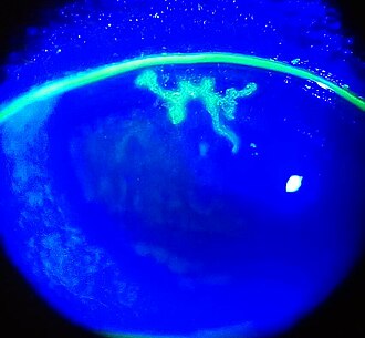
HSV dendritic keratitis — source media 1
IMAGE 01 · 330 × 305 px · Cached rendition

HSV dendritic keratitis

Viewed cached 330-pixel preview: an actual blue-light fluorescein clinical photograph showing a branching dendritic corneal ulcer. The source establishes the dendritic fluorescein pattern, but does not independently state HSV etiology.

Credit & reuse

Imrankabirhossain · CC BY-SA 4.0 · inherited_catalog_not_refetched

License record ↗ · Media ID MED-175efb76f00711593d46
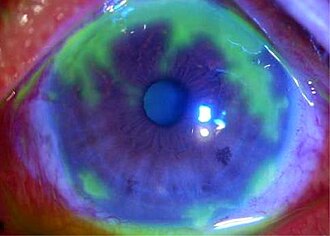
HSV dendritic keratitis — source media 2
IMAGE 02 · 330 × 236 px · Cached rendition

HSV dendritic keratitis

Viewed cached 330-pixel preview: an actual slit-lamp/fluorescein clinical eye photograph; the source explicitly identifies herpes-simplex geographic corneal ulcer. It supports HSV keratitis but shows a geographic ulcer rather than the requested classic dendritic branch.

Credit & reuse

Hee K Yang, Young K Han, Won R Wee, Jin H Lee and Ji W Kwon; Department of Ophthalmology, Seoul National University College of Medicine, Seoul, Korea · CC BY 2.5 · inherited_catalog_not_refetched

License record ↗ · Media ID MED-4544a99638798f7b9126