Skip to content
thecodexes.comIMAGE BANK
PLATE EYE-CRAO · 3 SOURCE FILES

Central retinal artery occlusion

Study the finding, then test the visual cue against the linked Review and Q-Book material.

View current live plate ↗
THE PLATE03 / 03
Central retinal artery occlusion fundus — source media 1
IMAGE 01 · 762 × 656 px · Cached rendition

Central retinal artery occlusion fundus

Fundus photo shows retinal whitening around a central cherry-red macular spot and attenuated vessels.

Credit & reuse

Sehreen Mumtaz, Claire Wilson, Prasanna Vibhute, Eric R Eggenberger, Florentina Berianu, and Andy Abril · CC BY 4.0 · source_rechecked_this_run

License record ↗ · Media ID MED-44d2d8fccff575b8bcef
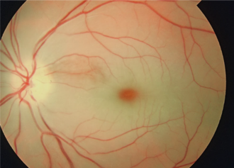
Central retinal artery occlusion — source media 3
IMAGE 03 · 330 × 246 px · Cached rendition

Central retinal artery occlusion

Source, credit & reuse

Source description identifies typical CRAO with a cherry-red spot, retinal edema, and narrowed vessels.

Achim Fieß, Ömer Cal, Stephan Kehrein, Sven Halstenberg, Inez Frisch, Ulrich Helmut Steinhorst · CC BY 2.0 · inherited_catalog_not_refetched

License record ↗ · Media ID MED-07c16633f7b53845d430