Skip to content
thecodexes.comIMAGE BANK
PLATE EYE-CRVO · 2 SOURCE FILES

Central retinal vein occlusion

Study the finding, then test the visual cue against the linked Review and Q-Book material.

View current live plate ↗
THE PLATE02 / 02
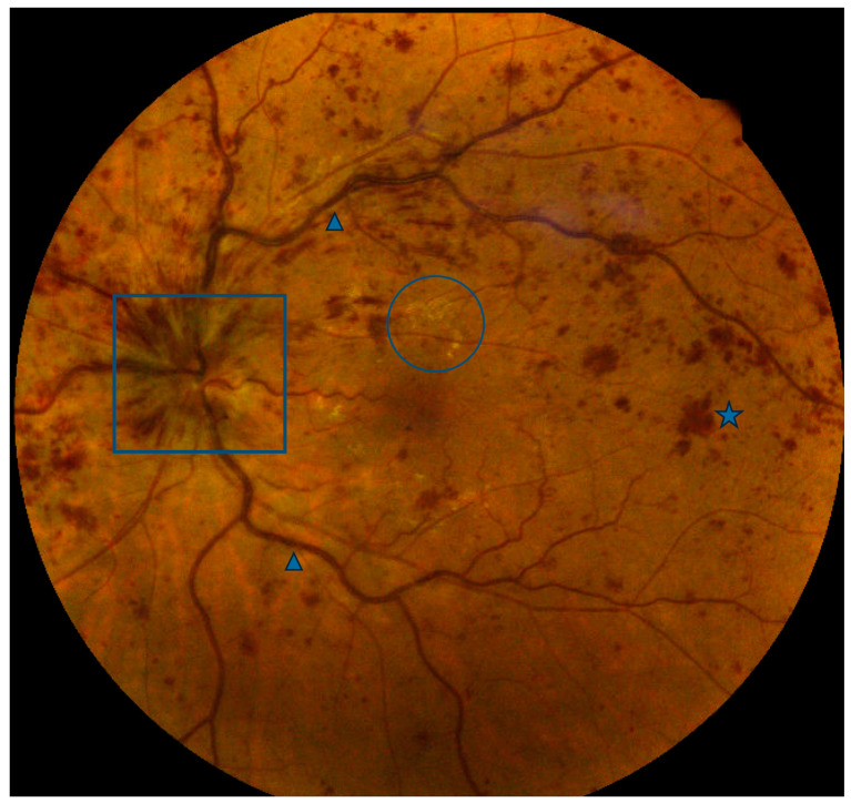
Central retinal vein occlusion fundus — source media 1
IMAGE 01 · 767 × 724 px · Cached rendition

Central retinal vein occlusion fundus

Fundus photo shows extensive intraretinal hemorrhages, dilated tortuous veins, and optic-disc edema.

Credit & reuse

Joseph Colcombe, Rusdeep Mundae, Alexis Kaiser, Jacques Bijon, and Yasha Modi · CC BY 4.0 · source_rechecked_this_run

License record ↗ · Media ID MED-1dad8867bcd39df66dc3
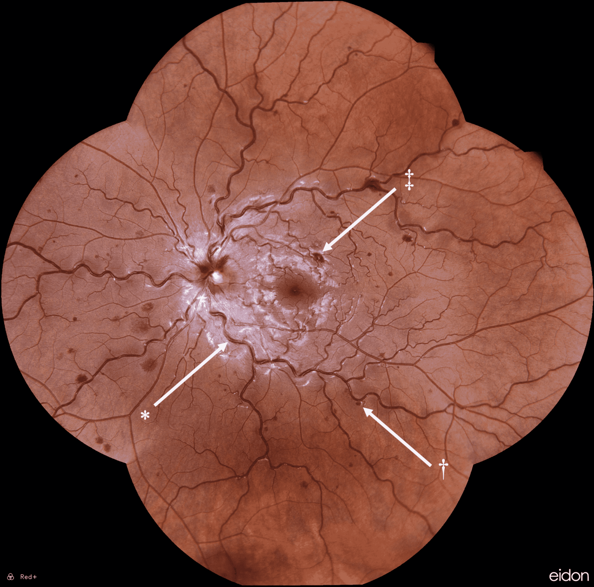
Central retinal vein occlusion — source media 2
IMAGE 02 · 1920 × 1898 px · Cached rendition

Central retinal vein occlusion

Source, credit & reuse

Source description identifies fundoscopy in CRVO with dilated tortuous veins and dot-blot hemorrhages; full blood-and-thunder appearance remains unconfirmed.

Alexander M Balinski, Rachel N Harvey, Ryan B Ko, Melanie M Smalley, Nathan E Cutler, and Maham T Siddiqi · CC BY 4.0 · inherited_catalog_not_refetched

License record ↗ · Media ID MED-1f1429b25614184438c3