Skip to content
thecodexes.comIMAGE BANK
PLATE CT-SUBDURAL · 3 SOURCE FILES

Subdural hematoma CT

Study the finding, then test the visual cue against the linked Review and Q-Book material.

View current live plate ↗
THE PLATE03 / 03
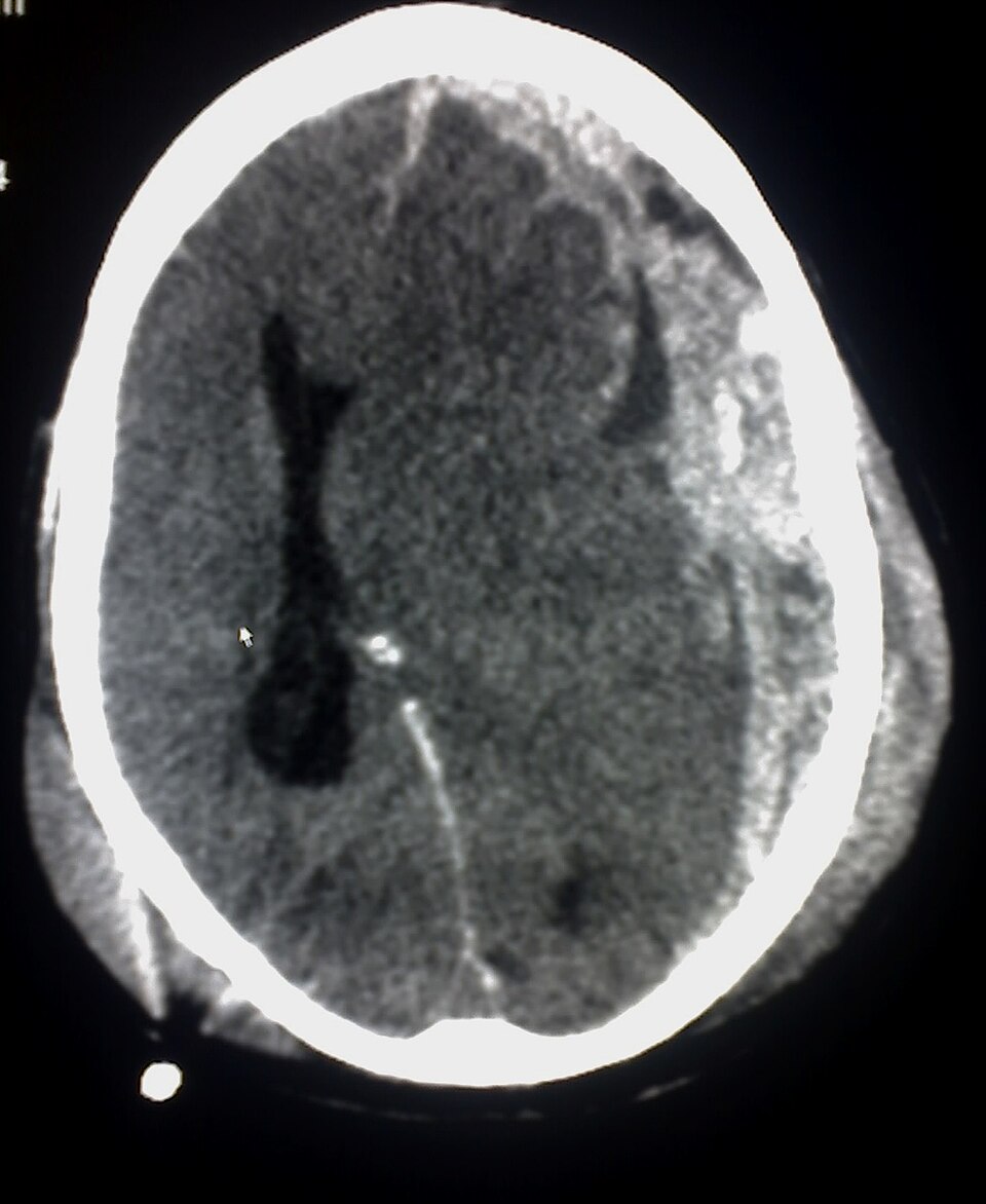
Subdural hematoma CT — source media 1
IMAGE 01 · 960 × 1172 px · Cached rendition

Subdural hematoma CT

Source, credit & reuse

Diagnosis and CT modality are explicit; crescentic shape, side, and midline shift are not separately confirmed in Source description.

Glitzy queen00 at English Wikipedia · Public domain · inherited_catalog_not_refetched

License record ↗ · Media ID MED-a03c800c5d03f076e5a8
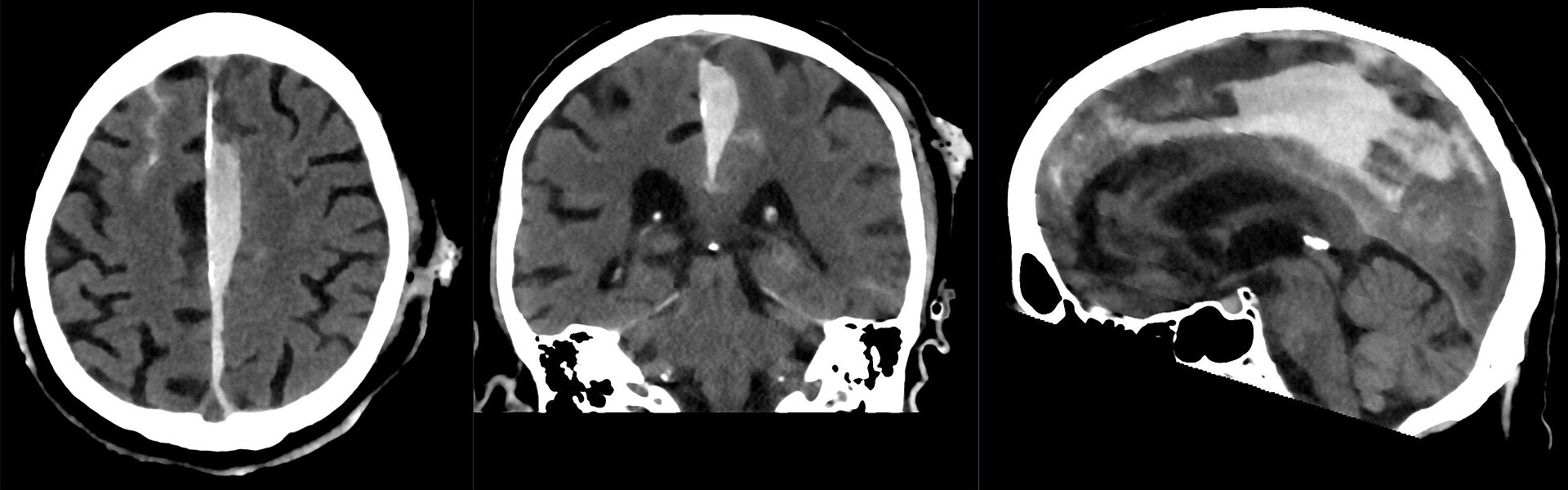
Subdural hematoma CT — source media 2
IMAGE 02 · 1920 × 600 px · Cached rendition

Subdural hematoma CT

Source, credit & reuse

Source description identifies CT of a parafalcine subdural hematoma; crescent shape and suture crossing remain unconfirmed.

Hellerhoff · CC BY-SA 4.0 · inherited_catalog_not_refetched

License record ↗ · Media ID MED-b41d8a85528ae033abb6

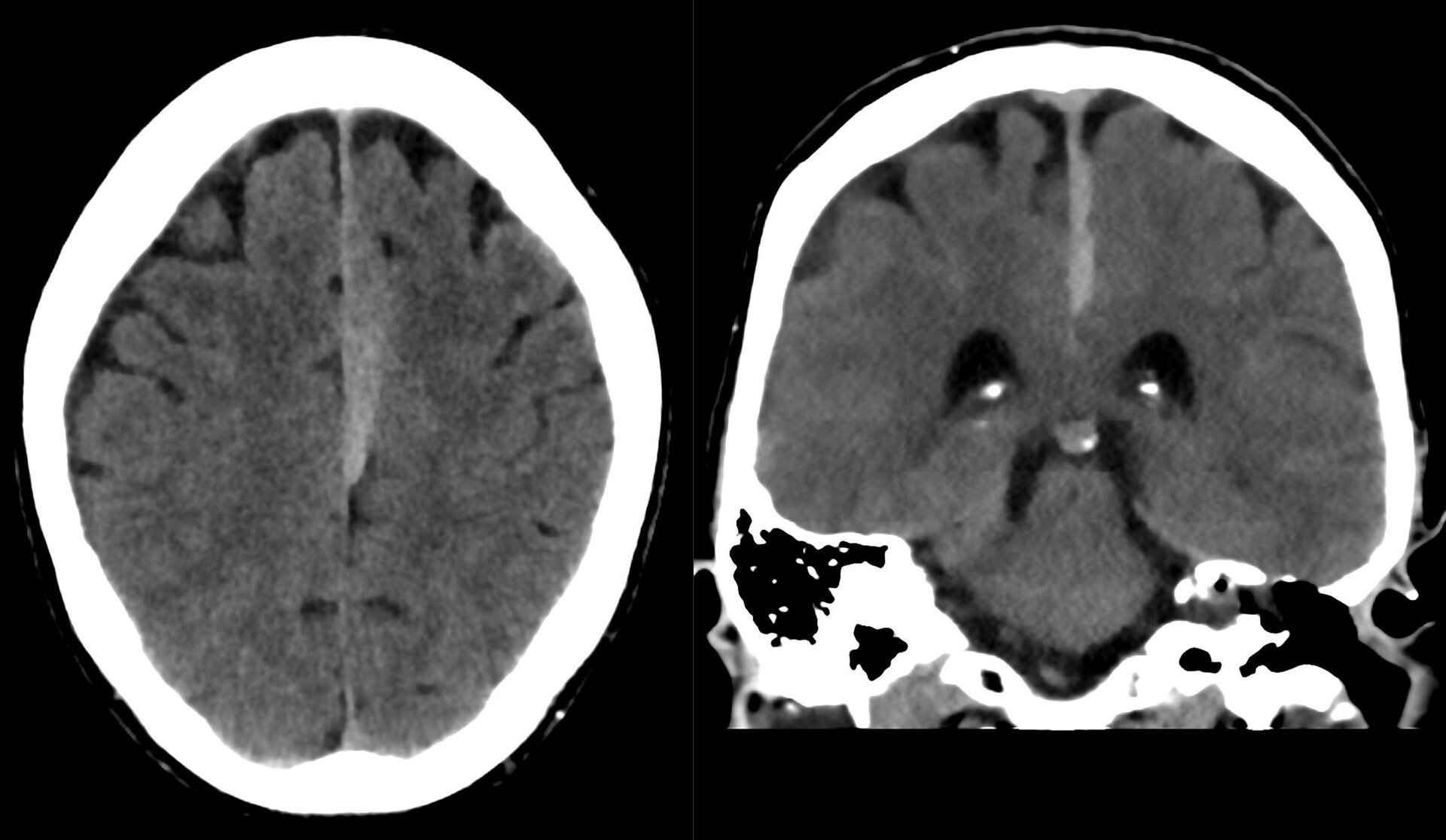
Subdural hematoma CT — source media 3
IMAGE 03 · 1920 × 1116 px · Cached rendition

Subdural hematoma CT

Diagnosis, left parafalcine location, and CT modality are explicit; this is not the classic convexity presentation.

Credit & reuse

Hellerhoff · CC BY-SA 4.0 · inherited_catalog_not_refetched

License record ↗ · Media ID MED-ffffdd4ad0966495f625