Skip to content
thecodexes.comIMAGE BANK
PLATE CT-ICH · 3 SOURCE FILES

Intracerebral hemorrhage CT

Study the finding, then test the visual cue against the linked Review and Q-Book material.

View current live plate ↗
THE PLATE03 / 03
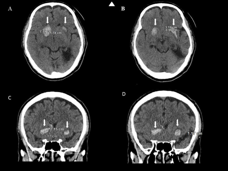
Bilateral basal ganglia hemorrhage — source media 1
IMAGE 01 · 800 × 600 px · Cached rendition

Bilateral basal ganglia hemorrhage

Noncontrast CT panels show bilateral hyperdense basal ganglia/lentiform parenchymal hemorrhages.

Credit & reuse

Tariq A Shaheed, Nicholas Glover, and Safwan Alboiny · CC BY 4.0 · source_rechecked_this_run

License record ↗ · Media ID MED-52d6c56f43b09629863b
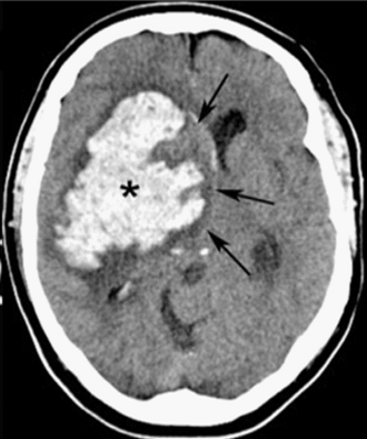
Intracerebral hemorrhage CT — source media 2
IMAGE 02 · 330 × 395 px · Cached rendition

Intracerebral hemorrhage CT

Diagnosis/location and CT modality are explicit; hypertensive etiology and precise putaminal involvement are unconfirmed. Lower-resolution fallback.

Credit & reuse

Shazia Mirza and Sankalp Gokhale · CC BY 4.0 · inherited_catalog_not_refetched

License record ↗ · Media ID MED-c652a08c9503ccbf1d89

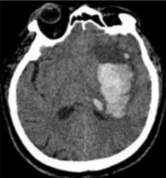
Intracerebral hemorrhage CT — source media 3
IMAGE 03 · 330 × 353 px · Cached rendition

Intracerebral hemorrhage CT

Diagnosis/location and CT modality are explicit; hypertensive etiology and putaminal sublocation are unconfirmed. Lower-resolution fallback.

Credit & reuse

Mats B. Dahlqvist et al. · CC BY 2.0 · inherited_catalog_not_refetched

License record ↗ · Media ID MED-e5576bb5edbf376b504b