Skip to content
thecodexes.comIMAGE BANK
PLATE CT-SAH · 4 SOURCE FILES

Subarachnoid hemorrhage CT

Study the finding, then test the visual cue against the linked Review and Q-Book material.

View current live plate ↗
THE PLATE04 / 04
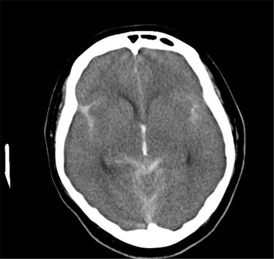
Large subarachnoid hemorrhage with hydrocephalus — source media 1
IMAGE 01 · 400 × 378 px · Cached rendition

Large subarachnoid hemorrhage with hydrocephalus

Axial noncontrast head CT shows hyperdense subarachnoid blood in basal cisterns/sylvian spaces.

Credit & reuse

Yukari Wakabayashi and Goshi Takahashi · CC BY 4.0 · source_rechecked_this_run

License record ↗ · Media ID MED-31e6873f277fc572b68b
Subarachnoid hemorrhage CT — source media 2
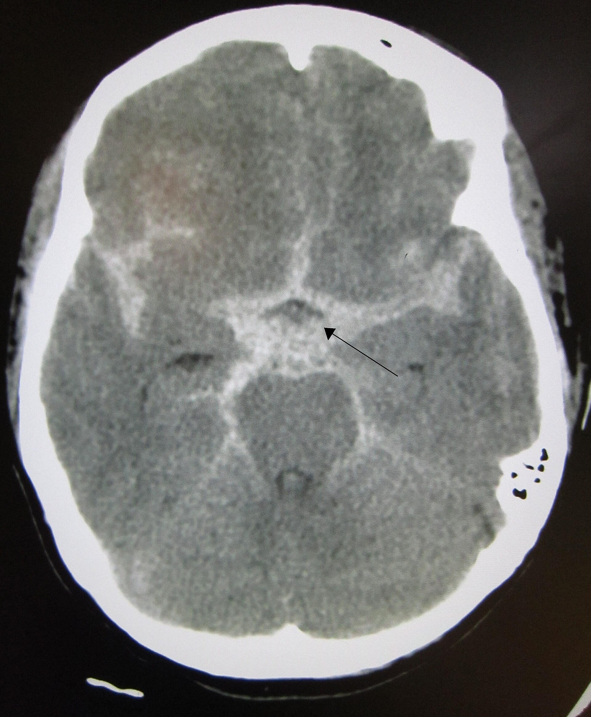
IMAGE 02 · 1920 × 2331 px · Cached rendition

Subarachnoid hemorrhage CT

Diagnosis and CT modality/category are explicit; basal-cistern distribution and aneurysmal cause are unconfirmed.

Credit & reuse

James Heilman, MD · CC BY-SA 3.0 · inherited_catalog_not_refetched

License record ↗ · Media ID MED-31be7f2a56e1ad21ca79

Subarachnoid hemorrhage CT — source media 3
IMAGE 03 · 1920 × 2082 px · Cached rendition

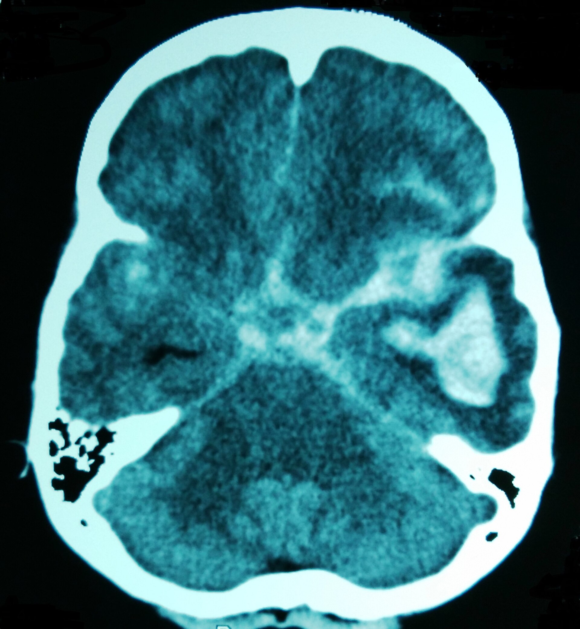
Subarachnoid hemorrhage CT

Diagnosis and CT modality are explicit; concomitant temporal hemorrhage may distract and basal-cistern distribution is unconfirmed.

Credit & reuse

Cerevisae · CC BY-SA 4.0 · inherited_catalog_not_refetched

License record ↗ · Media ID MED-5cbb854d0c2ff88540bc

Subarachnoid hemorrhage CT — source media 4
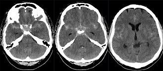
IMAGE 04 · 330 × 145 px · Cached rendition

Subarachnoid hemorrhage CT

Source, credit & reuse

Source description identifies axial CT of basal subarachnoid hemorrhage; sylvian-fissure involvement remains unconfirmed.

Hellerhoff · CC BY-SA 4.0 · inherited_catalog_not_refetched

License record ↗ · Media ID MED-4dc023762b9ce48b5e4a