Skip to content
thecodexes.comIMAGE BANK
PLATE US-GALLSTONES · 3 SOURCE FILES

Acute cholecystitis ultrasound

Study the finding, then test the visual cue against the linked Review and Q-Book material.

View current live plate ↗
THE PLATE03 / 03
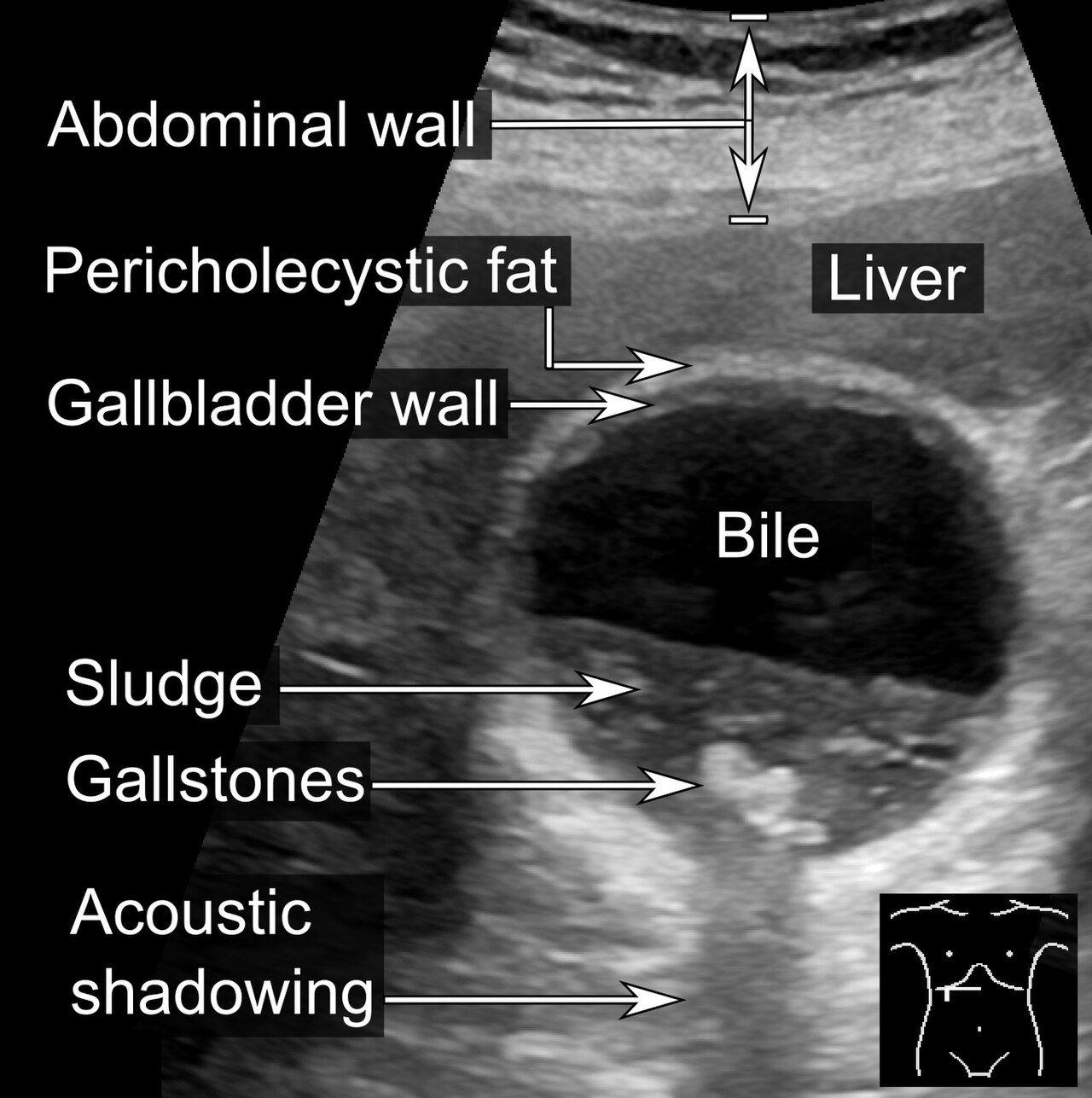
Acute cholecystitis ultrasound — source media 1
IMAGE 01 · 330 × 225 px · Cached rendition

Acute cholecystitis ultrasound

Source, credit & reuse

Source description identifies acute cholecystitis on ultrasound; stone, acoustic shadowing, wall thickening, and fluid remain unconfirmed.

Cerevisae · CC BY-SA 4.0 · inherited_catalog_not_refetched

License record ↗ · Media ID MED-191a448171940d489edb
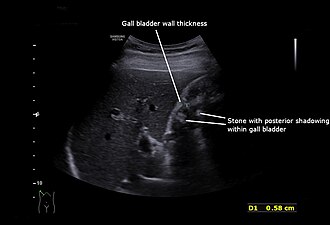
Acute cholecystitis ultrasound — source media 2
IMAGE 02 · 1920 × 2560 px · Cached rendition

Acute cholecystitis ultrasound

Diagnosis and ultrasound modality are explicit; posterior acoustic shadowing and mobility are unconfirmed.

Credit & reuse

Cerevisae · CC BY-SA 4.0 · inherited_catalog_not_refetched

License record ↗ · Media ID MED-b95d348c2b17ba81175e

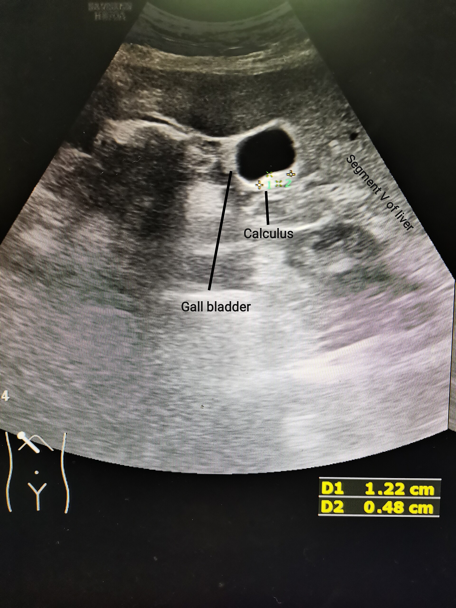
Acute cholecystitis ultrasound — source media 3
IMAGE 03 · 1280 × 1287 px · Cached rendition

Acute cholecystitis ultrasound

Diagnosis and ultrasound modality are explicit; coexisting sludge may distract and exact acoustic-shadowing detail is unconfirmed.

Credit & reuse

Mikael Häggström , M.D. Author info - Reusing images - Conflicts of interest: None Mikael Häggström , M.D. Consent note : Written informed consent was obtained from the individual, including online publication. · CC0 · inherited_catalog_not_refetched

License record ↗ · Media ID MED-1c4cb78ab39c3d13cf48