Skip to content
thecodexes.comIMAGE BANK
PLATE US-B11-042 · 2 SOURCE FILES

Absent or markedly decreased color Doppler flow to the affected testis with an…

Study the finding, then test the visual cue against the linked Review and Q-Book material.

View current live plate ↗
THE PLATE02 / 02
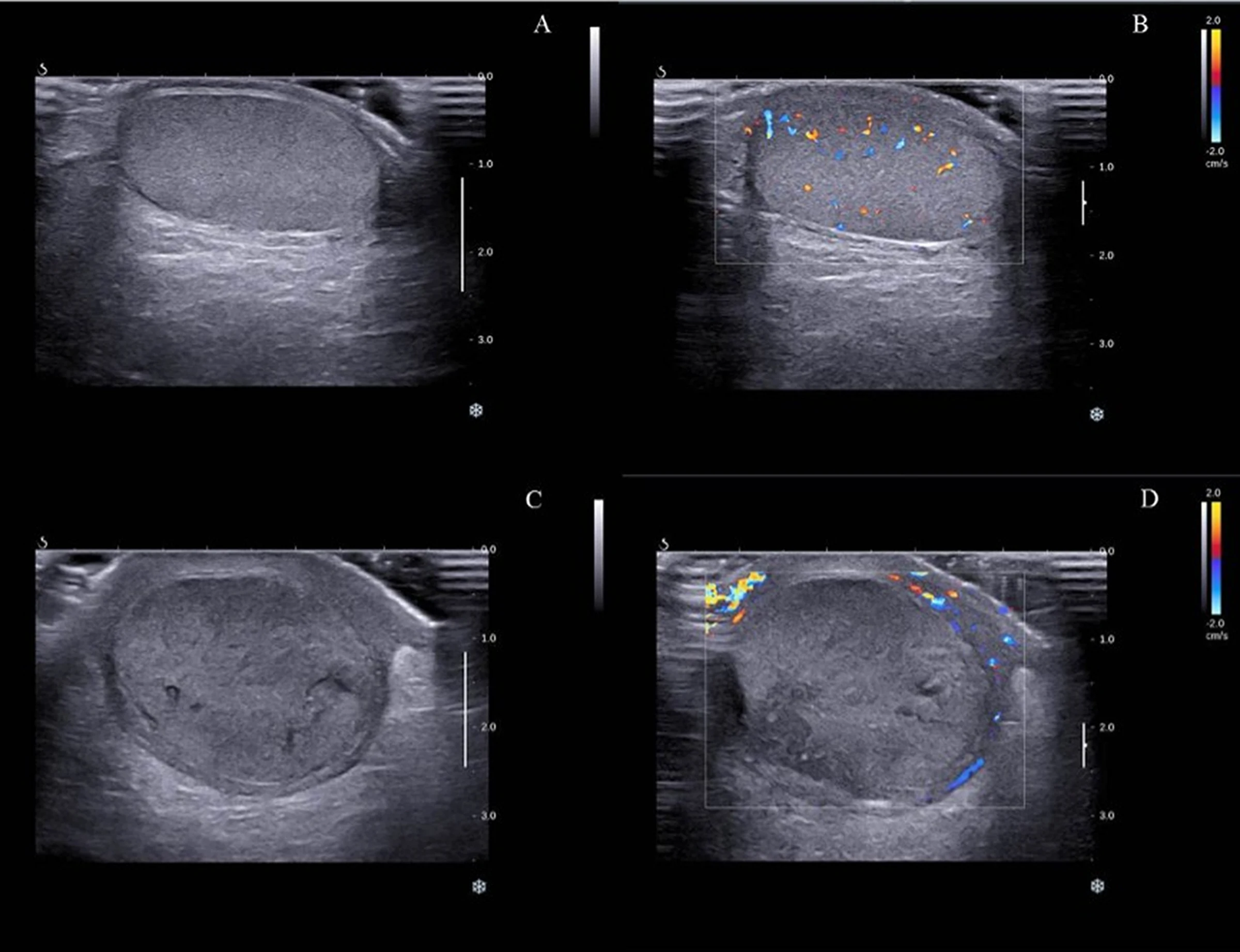
Normal versus torsed testis on gray-scale and color Doppler — source media 1
IMAGE 01 · 2417 × 1856 px · Browser preview; TIFF original available

Normal versus torsed testis on gray-scale and color Doppler

Four-panel original figure compares gray-scale and color Doppler of a normal testis with an enlarged torsed testis lacking detectable intratesticular color flow.

Credit & reuse

Zhenxing Sun, Mingxing Xie, Feixiang Xiang, Yue Song, Cheng Yu, Yanrong Zhang, Sachin Ramdhany, and Jing Wang · CC BY 4.0 · source_rechecked_this_run

License record ↗ · Media ID MED-de03108f5eaed95104f2
Absent right testicular flow versus left — source media 2
IMAGE 02 · 705 × 647 px · Cached rendition

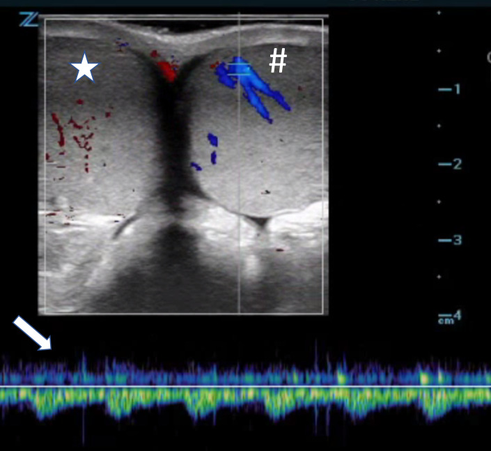
Absent right testicular flow versus left

Color Doppler shows absent/minimal right intratesticular color signal versus preserved left-sided flow; spectral waveform accompanies the comparison.

Credit & reuse

Rahul V Nene, Rachna Subramony, Michael Macias, Colleen Campbell, and Amir Aminlari · CC BY 4.0 · source_rechecked_this_run

License record ↗ · Media ID MED-234adb41dbcf17655759