Skip to content
thecodexes.comIMAGE BANK
PLATE SMEAR-SCHISTOCYTES · 3 SOURCE FILES

Schistocytes blood smear

Study the finding, then test the visual cue against the linked Review and Q-Book material.

View current live plate ↗
THE PLATE03 / 03
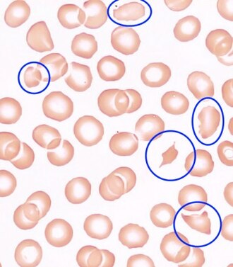
Schistocytes blood smear — source media 1
IMAGE 01 · 330 × 377 px · Cached rendition

Schistocytes blood smear

Source, credit & reuse

Source description identifies a blood smear with typical schistocytes in TTP; helmet-cell morphology remains unconfirmed.

Central Hematology Laboratory Hemostasis Research Laboratory Bern University Hospital & University of Bern · CC BY-SA 4.0 · inherited_catalog_not_refetched

License record ↗ · Media ID MED-19a308c300bf29db30bb
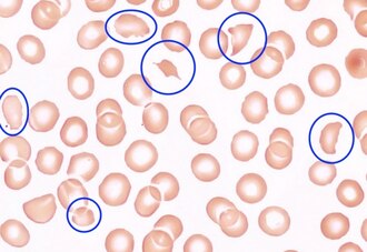
Schistocytes blood smear — source media 2
IMAGE 02 · 330 × 227 px · Cached rendition

Schistocytes blood smear

Source, credit & reuse

Source description identifies a blood smear with typical schistocytes in TTP; helmet-cell morphology remains unconfirmed.

Central Hematology Laboratory Hemostasis Research Laboratory Bern University Hospital & University of Bern · CC BY-SA 4.0 · inherited_catalog_not_refetched

License record ↗ · Media ID MED-314b41dfa6b57f64aae3

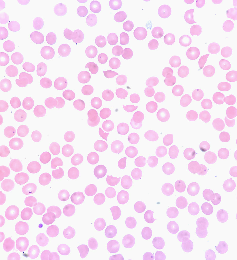
Schistocytes blood smear — source media 3
IMAGE 03 · 936 × 1024 px · Cached rendition

Schistocytes blood smear

Source title/caption identifies the requested cellular finding; preview visually confirms a stained smear field. Source title and description support the requested cellular finding. Exact specimen provenance and every requested subfeature require clinical review.

Credit & reuse

Ed Uthman · CC BY 4.0 · inherited_catalog_not_refetched

License record ↗ · Media ID MED-824849bd64ae0e18a356