Skip to content
thecodexes.comIMAGE BANK
PLATE SMEAR-REED-STERNBERG · 3 SOURCE FILES

Reed Sternberg cell

Study the finding, then test the visual cue against the linked Review and Q-Book material.

View current live plate ↗
THE PLATE03 / 03
Reed Sternberg cell — source media 1
IMAGE 01 · 1920 × 1440 px · Cached rendition

Reed Sternberg cell

Source, credit & reuse

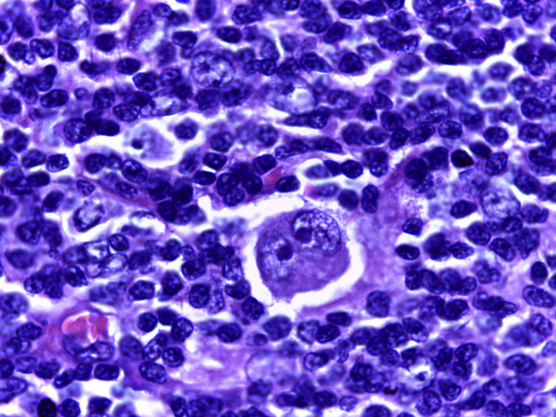
Source description identifies lymph-node micrograph in Hodgkin lymphoma with a characteristic binucleate Reed-Sternberg cell.

Calicut Medical College · CC BY-SA 4.0 · inherited_catalog_not_refetched

License record ↗ · Media ID MED-59cc5af9c9f0d8564e14
Reed Sternberg cell — source media 2
IMAGE 02 · 1024 × 840 px · Cached rendition

Reed Sternberg cell

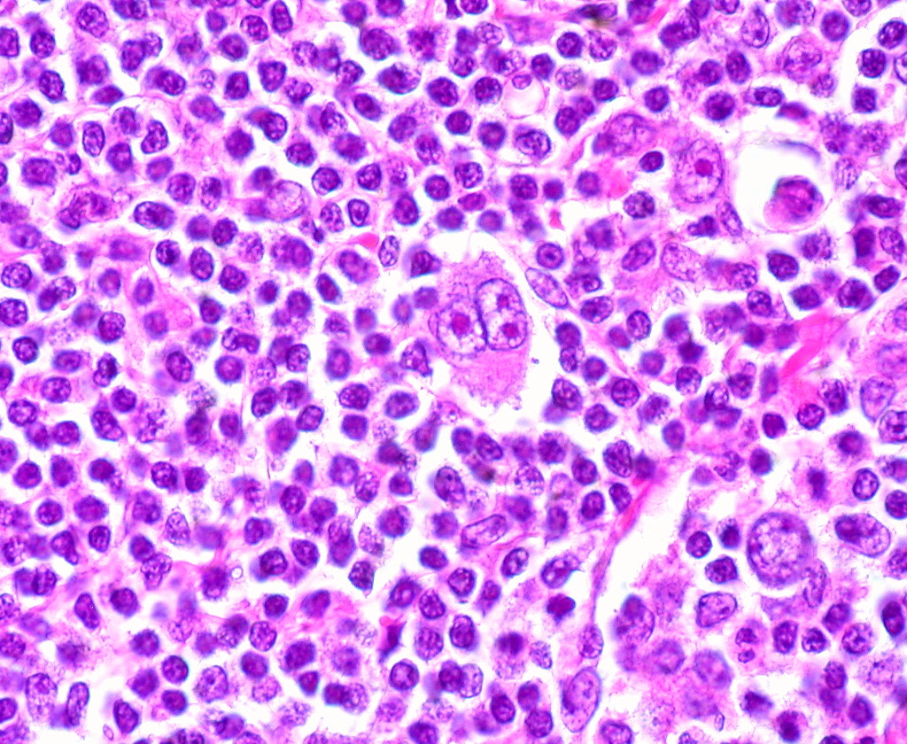
Source title/caption identifies the requested cellular finding; preview visually confirms a histology section. Source title and description support the requested cellular finding. Exact specimen provenance and every requested subfeature require clinical review.

Credit & reuse

Ed Uthman · CC BY 4.0 · inherited_catalog_not_refetched

License record ↗ · Media ID MED-92c2a00fddcec16859d2

Reed Sternberg cell — source media 3
IMAGE 03 · 1024 × 768 px · Cached rendition

Reed Sternberg cell

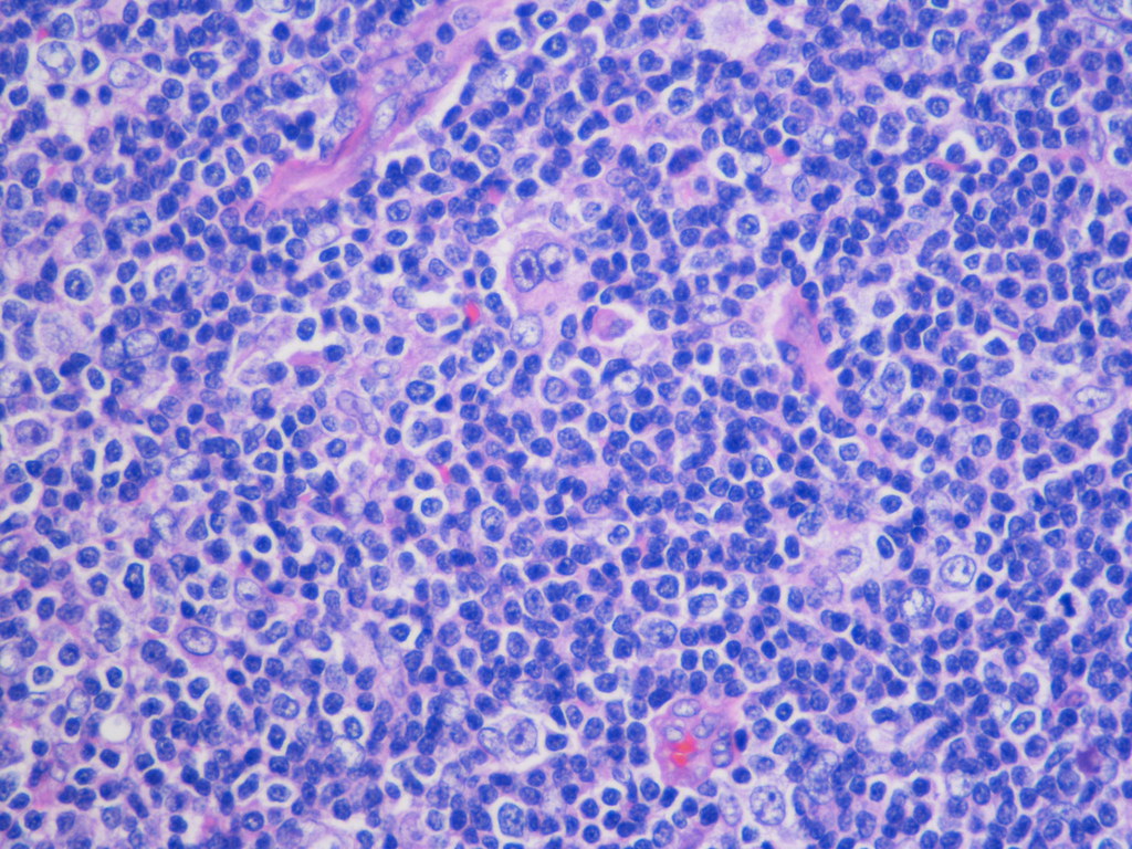
Source title/caption identifies the requested cellular finding; preview visually confirms a histology section. Source title and description support the requested cellular finding. Exact specimen provenance and every requested subfeature require clinical review.

Credit & reuse

cnicholsonpath · CC BY 2.0 · inherited_catalog_not_refetched

License record ↗ · Media ID MED-fa3194093f1d56977c59