Skip to content
thecodexes.comIMAGE BANK
PLATE SMEAR-CLL-SMUDGE · 3 SOURCE FILES

CLL smudge cells

Study the finding, then test the visual cue against the linked Review and Q-Book material.

View current live plate ↗
THE PLATE03 / 03
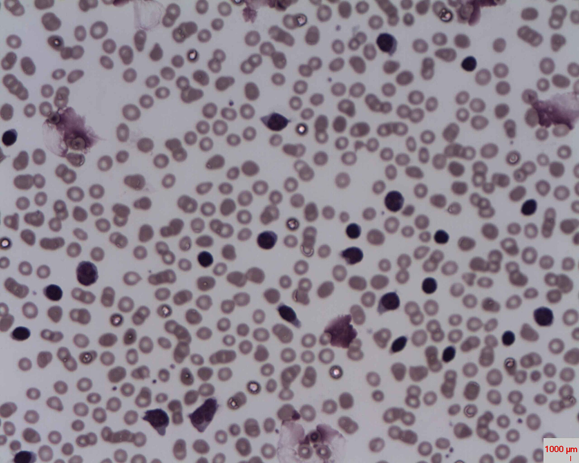
CLL smudge cells — source media 1
IMAGE 01 · 1920 × 1536 px · Cached rendition

CLL smudge cells

Source, credit & reuse

Source description identifies a CLL blood smear with marked lymphocytosis and smear/smudge cells; exact lymphocyte morphology remains unconfirmed.

Erhabor Osaro (Associate Professor) · CC BY-SA 3.0 · inherited_catalog_not_refetched

License record ↗ · Media ID MED-ad4dfffc9bdbca928752
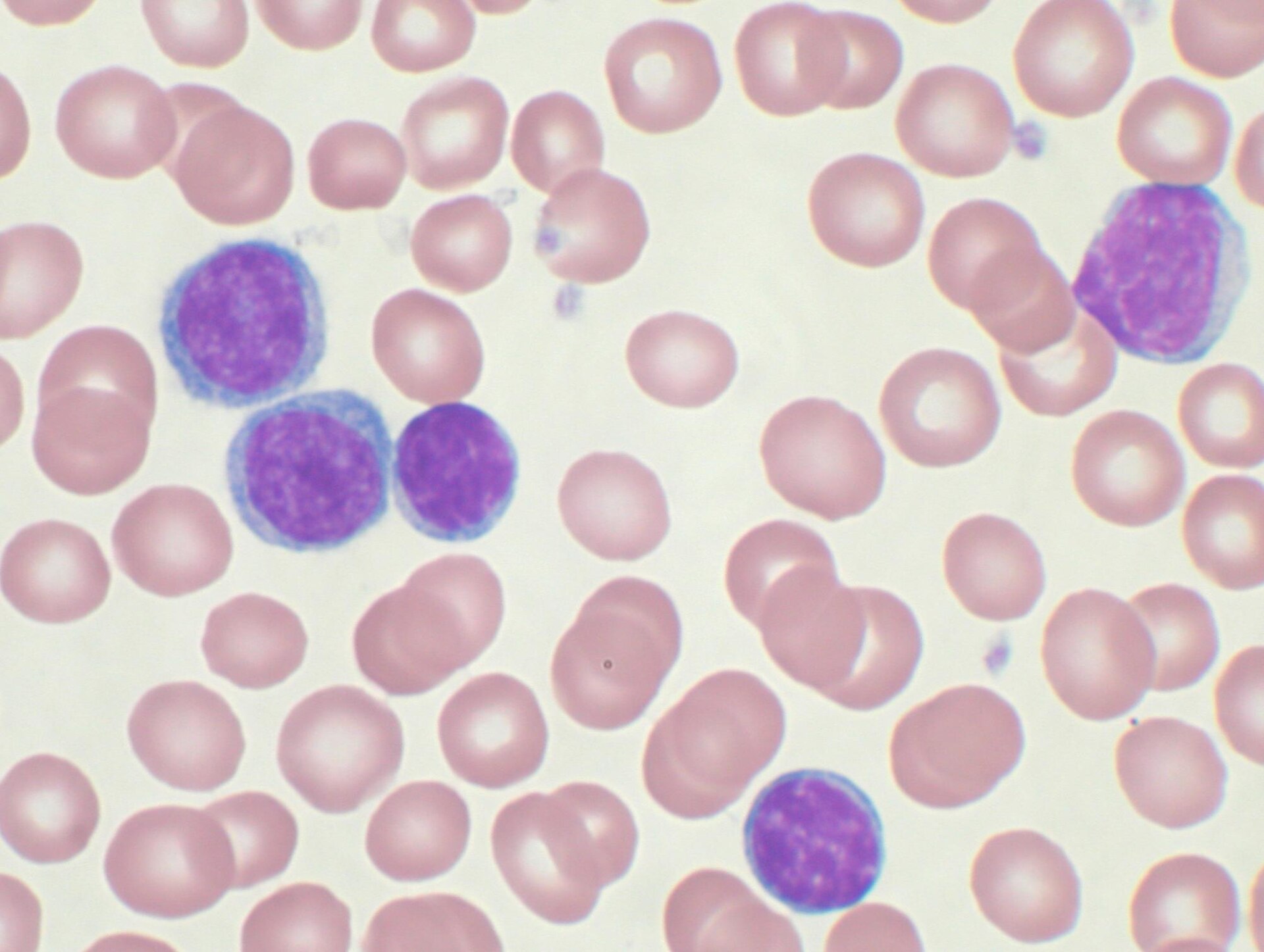
CLL smudge cells — source media 2
IMAGE 02 · 1920 × 1447 px · Cached rendition

CLL smudge cells

Source, credit & reuse

Source description identifies a Wright-stained peripheral blood smear of CLL with mature-appearing lymphocytes; smudge cells remain unconfirmed.

Mary Ann Thompson ( VashiDonsk at en.wikipedia ) · CC BY-SA 3.0 · inherited_catalog_not_refetched

License record ↗ · Media ID MED-000a4abd938b06221cd2

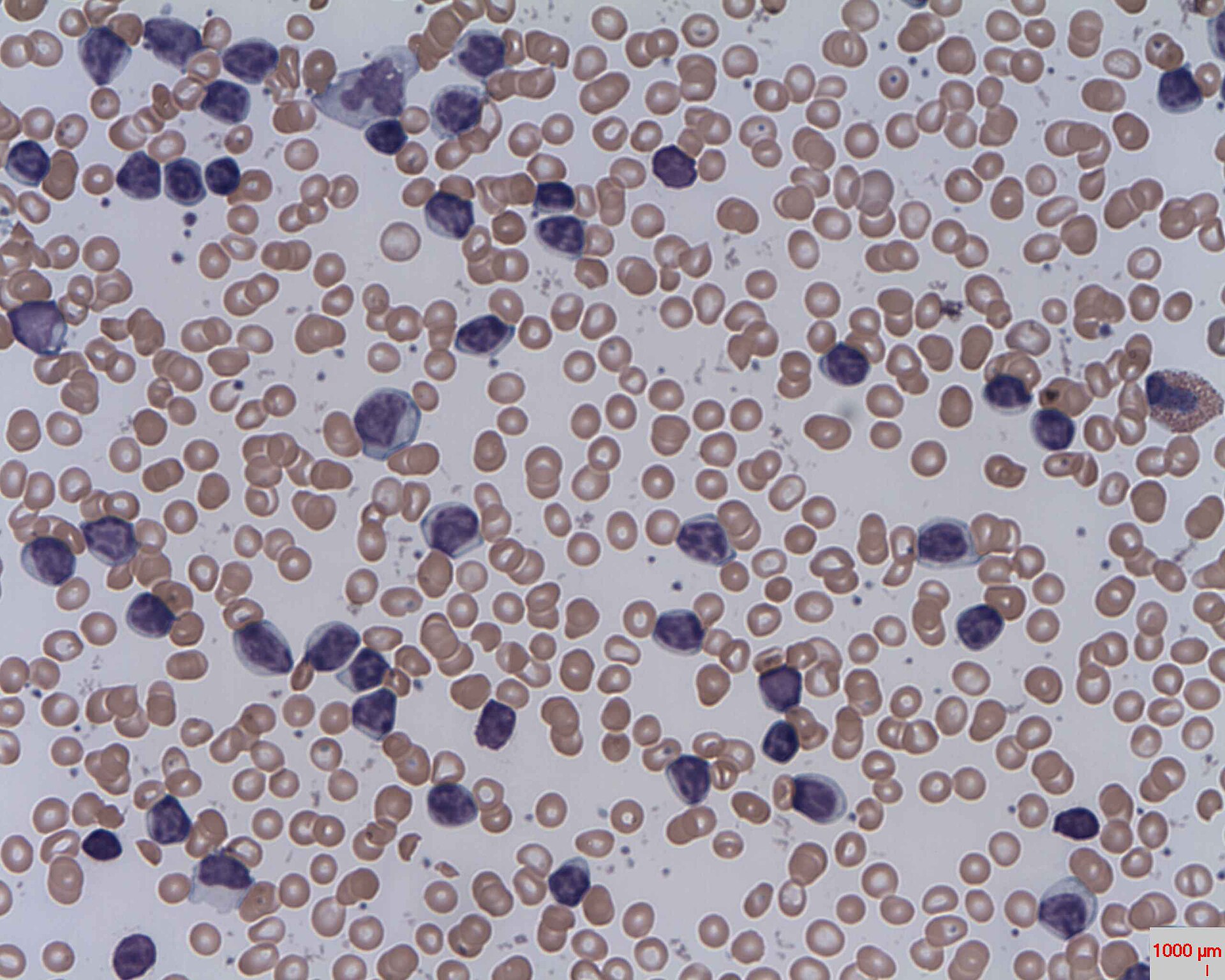
CLL smudge cells — source media 3
IMAGE 03 · 1920 × 1536 px · Cached rendition

CLL smudge cells

Source, credit & reuse

Source description identifies a CLL blood smear with marked lymphocytosis and smear/smudge cells; exact lymphocyte morphology remains unconfirmed.

Prof. Erhabor Osaro · CC BY-SA 4.0 · inherited_catalog_not_refetched

License record ↗ · Media ID MED-a77378175745fb7ee328