Skip to content
thecodexes.comIMAGE BANK
PLATE SKIN-ZOSTER · 3 SOURCE FILES

Herpes zoster

Study the finding, then test the visual cue against the linked Review and Q-Book material.

View current live plate ↗
THE PLATE03 / 03
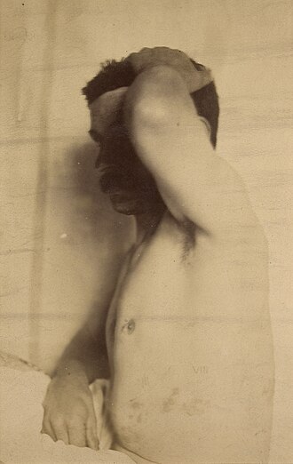
Herpes zoster — source media 1
IMAGE 01 · 330 × 522 px · Cached rendition

Herpes zoster

Viewed cached 330-pixel preview: a historical black-and-white clinical photograph of a unilateral flank eruption; the source explicitly identifies herpes zoster. Lesion detail is limited by the archival contrast.

Credit & reuse

Godart, Thomas · CC BY 4.0 · inherited_catalog_not_refetched

License record ↗ · Media ID MED-7b4ba77ac98f6096e804
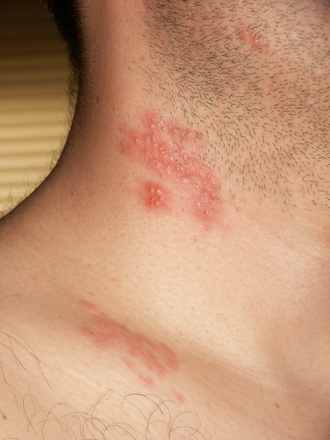
Herpes zoster — source media 2
IMAGE 02 · 330 × 440 px · Cached rendition

Herpes zoster

Viewed cached 330-pixel preview: an actual clinical photograph showing grouped erythematous lesions on one side of the neck/shoulder; the source explicitly identifies zoster blisters.

Credit & reuse

No machine-readable author provided. Gentgeen assumed (based on copyright claims). · CC BY-SA 3.0 · inherited_catalog_not_refetched

License record ↗ · Media ID MED-b9f221e2f4414ed343d2

Herpes zoster — source media 3
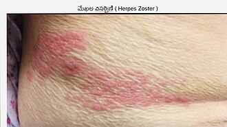
IMAGE 03 · 330 × 185 px · Cached rendition

Herpes zoster

Viewed cached 330-pixel preview: an actual clinical photograph showing a unilateral band of clustered erythematous lesions on the lower trunk; the source explicitly identifies herpes zoster. The image includes a text header.

Credit & reuse

డా. గన్నవరపు నరసింహమూర్తి · CC0 1.0 · inherited_catalog_not_refetched

License record ↗ · Media ID MED-47b2502c9655af29387f