Skip to content
thecodexes.comIMAGE BANK
PLATE MRI-MS · 3 SOURCE FILES

Multiple sclerosis MRI

Study the finding, then test the visual cue against the linked Review and Q-Book material.

View current live plate ↗
THE PLATE03 / 03
Multiple sclerosis MRI — source media 1
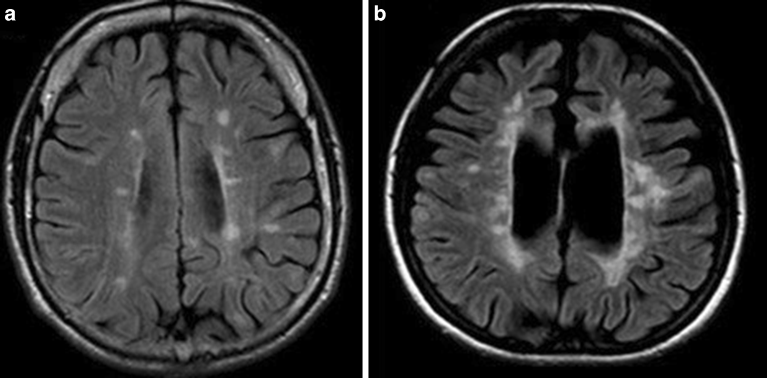
IMAGE 01 · 1524 × 751 px · Cached rendition

Multiple sclerosis MRI

Exact MRI and multiple-sclerosis match. The caption explicitly identifies periventricular, deep, and juxtacortical white-matter lesions. These are PPMS examples; they should not be presented as unique to PPMS or as diagnostic in isolation. Axial views; these do not demonstrate the requested sagittal Dawson-finger pattern.

Credit & reuse

Małgorzata Siger · CC BY 4.0 · inherited_catalog_not_refetched

License record ↗ · Media ID MED-c225748e68f5b4ef3e08
Multiple sclerosis MRI — source media 2
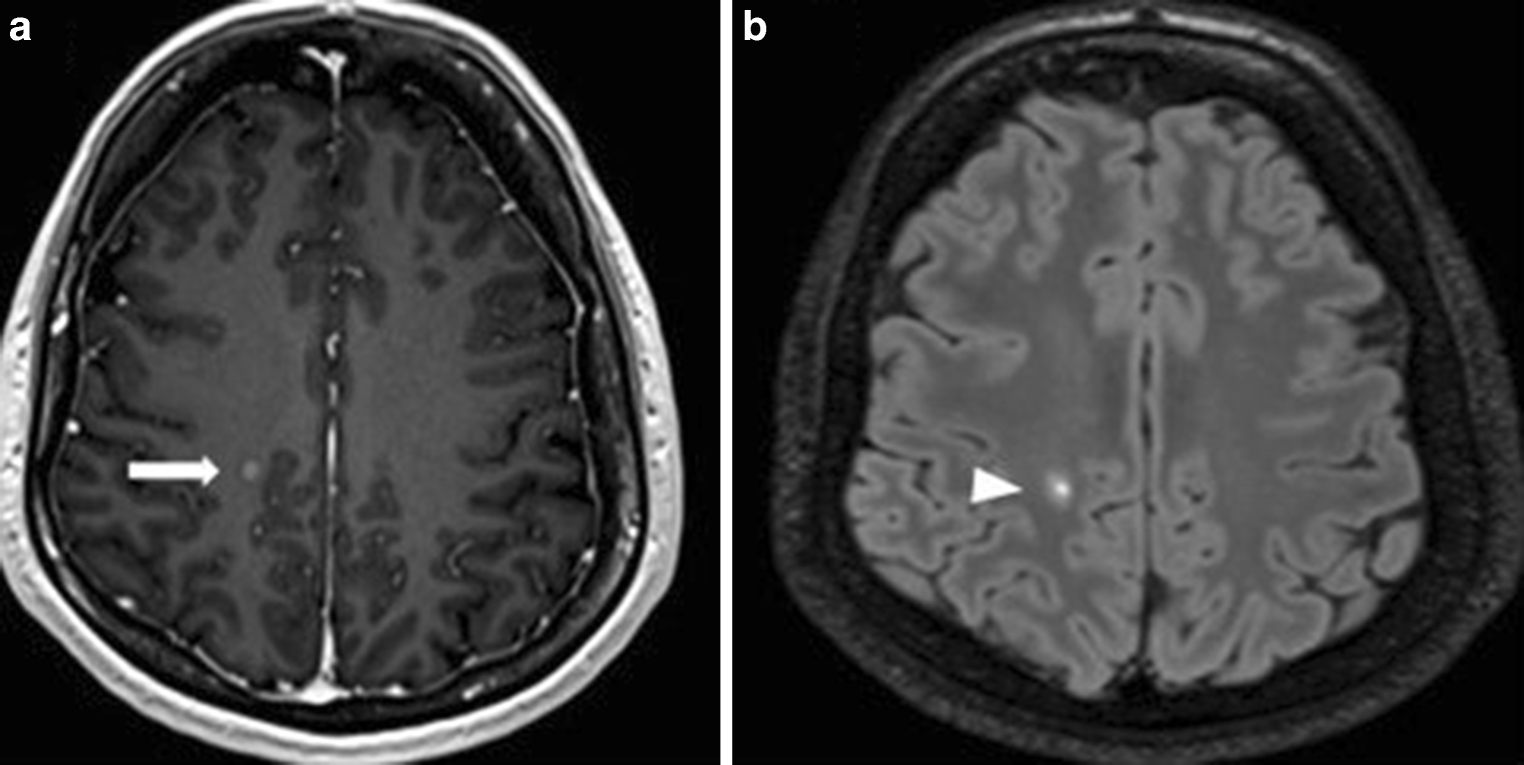
IMAGE 02 · 1524 × 765 px · Cached rendition

Multiple sclerosis MRI

Exact MRI and multiple-sclerosis match. The paired post-contrast T1 and FLAIR panels document an active enhancing lesion in a PPMS patient; enhancement is not present in every MS lesion. Axial views; these do not demonstrate the requested sagittal Dawson-finger pattern.

Credit & reuse

Małgorzata Siger · CC BY 4.0 · inherited_catalog_not_refetched

License record ↗ · Media ID MED-dd6fb36bb9c08ab8d2f5

Multiple sclerosis MRI — source media 3
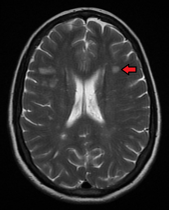
IMAGE 03 · 330 × 411 px · Cached rendition

Multiple sclerosis MRI

Source, credit & reuse

Source description identifies multiple sclerosis on MRI; sagittal FLAIR sequence and Dawson-finger morphology remain unconfirmed.

James Heilman, MD · CC BY-SA 4.0 · inherited_catalog_not_refetched

License record ↗ · Media ID MED-f74ecb6003cc5bde620e