IMAGE 01 · 1920 × 1280 px · Cached rendition
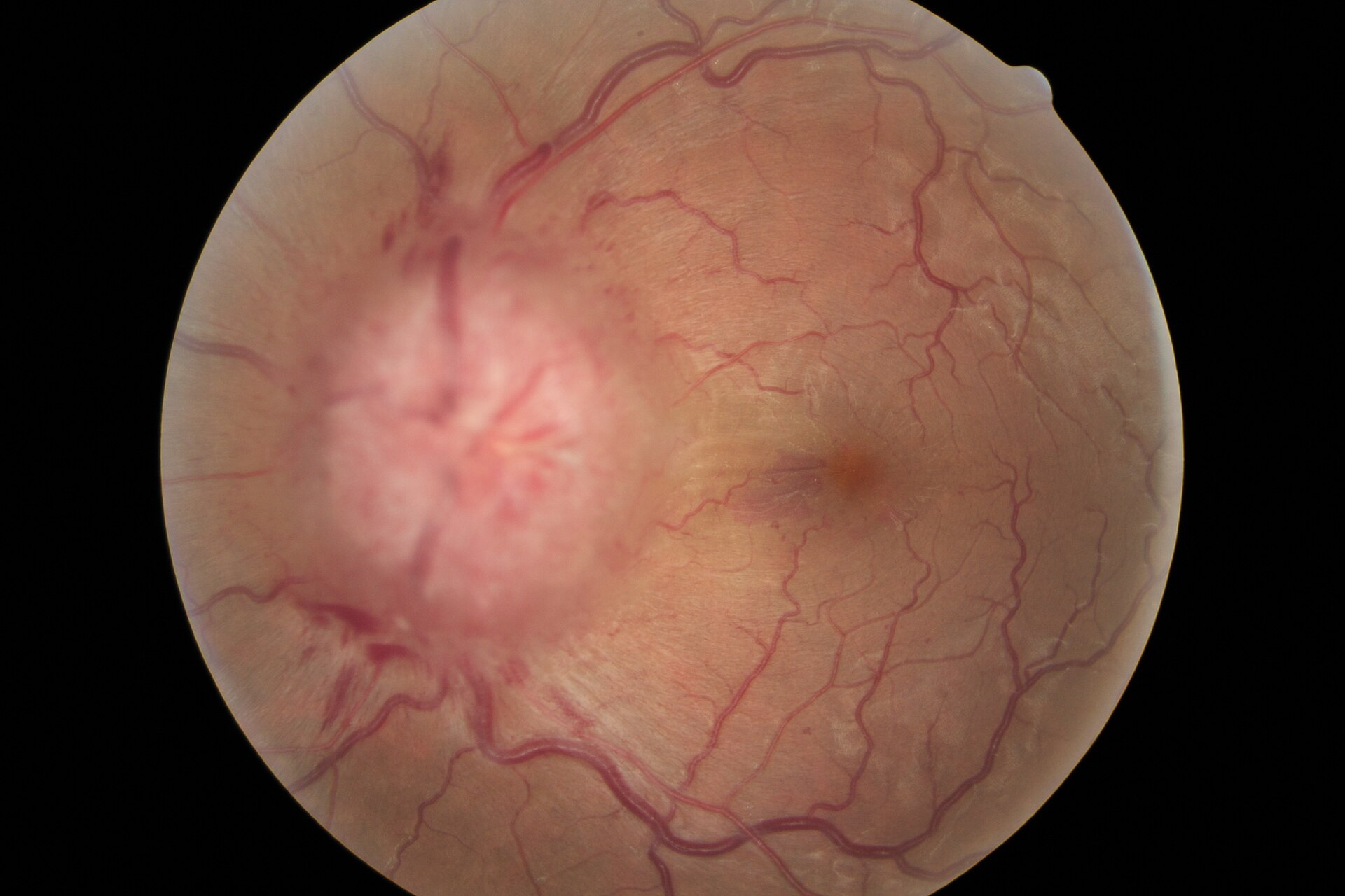
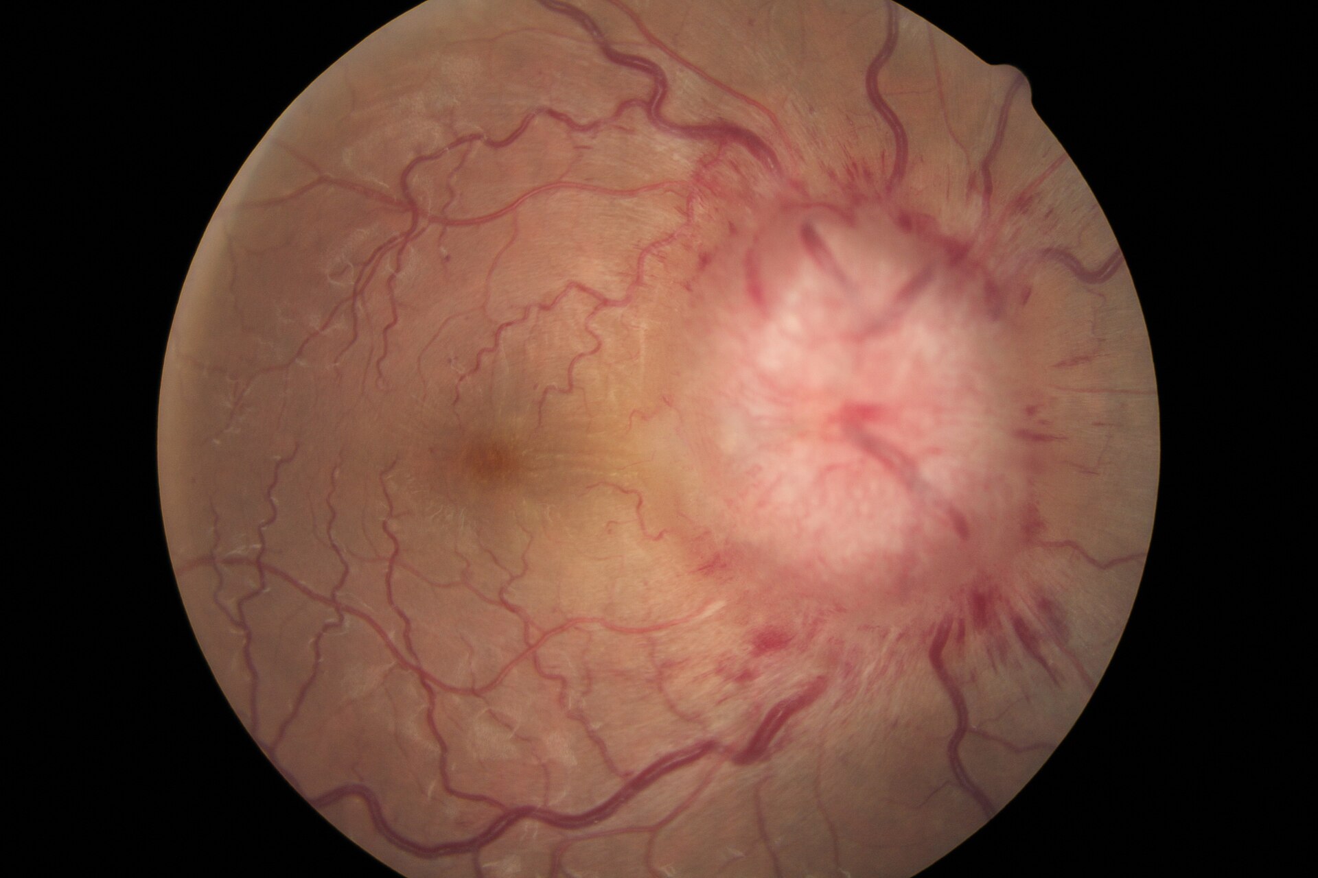
Papilledema fundus
Source caption and license checked.
Credit & reuse
DocSee04 · CC BY 4.0 · inherited_catalog_not_refetched
License record ↗ · Media ID MED-215f92df1bba9605b76e
Study the finding, then test the visual cue against the linked Review and Q-Book material.
Source caption and license checked.
DocSee04 · CC BY 4.0 · inherited_catalog_not_refetched
License record ↗ · Media ID MED-215f92df1bba9605b76e
Source caption and license checked.
DocSee04 · CC BY 4.0 · inherited_catalog_not_refetched
License record ↗ · Media ID MED-b114cca5ab74e0d58df7
Viewed cached 330-pixel preview: an actual retinal fundus photograph; the source explicitly identifies severe papilloedema. The small preview confirms the fundus modality, while exact hemorrhage and vessel-obscuration detail should be assessed from the native image.
Shveta Bansal , Timothy Dabbs and Vernon Long · CC BY 2.0 · inherited_catalog_not_refetched
License record ↗ · Media ID MED-1cb7281914e61113512d