Skip to content
thecodexes.comIMAGE BANK
PLATE EYE-KAYSER-FLEISCHER · 3 SOURCE FILES

Kayser Fleischer ring

Study the finding, then test the visual cue against the linked Review and Q-Book material.

View current live plate ↗
THE PLATE03 / 03
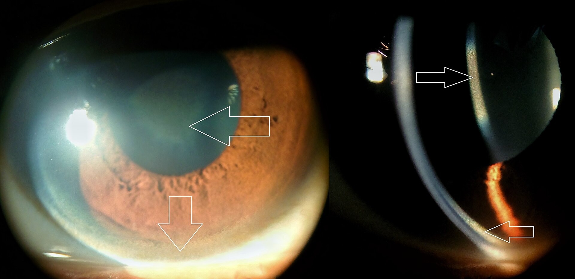
Kayser Fleischer ring — source media 1
IMAGE 01 · 1920 × 934 px · Cached rendition

Kayser Fleischer ring

Viewed cached thumbnail: a slit-lamp clinical-eye composite shows a peripheral Kayser-Fleischer ring and a sunflower cataract; the source description explicitly identifies copper deposition. The composite includes both findings, so the ring panel should be identified when displayed.

Credit & reuse

Imrankabirhossain · CC BY-SA 4.0 · inherited_catalog_not_refetched

License record ↗ · Media ID MED-47bbcf828b309b931f66
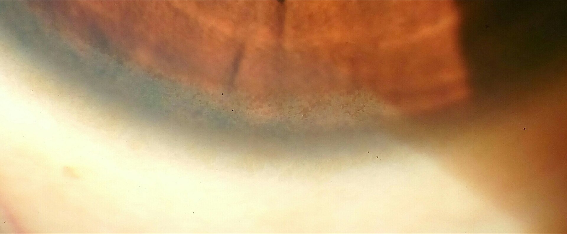
Kayser Fleischer ring — source media 2
IMAGE 02 · 1920 × 794 px · Cached rendition

Kayser Fleischer ring

Viewed cached thumbnail: an actual close-up clinical/slit-lamp photograph of the corneal limbus; the source identifies a child with Wilson disease and labels the file as a Kayser-Fleischer ring. Only a segment of the limbus is shown.

Credit & reuse

Imrankabirhossain · CC BY-SA 4.0 · inherited_catalog_not_refetched

License record ↗ · Media ID MED-db3e0268a887791b5823

Kayser Fleischer ring — source media 3
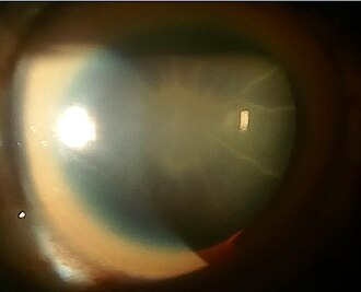
IMAGE 03 · 330 × 267 px · Cached rendition

Kayser Fleischer ring

Viewed cached 330-pixel preview: an actual slit-lamp eye photograph; the source explicitly identifies a thick Kayser-Fleischer ring together with a sunflower cataract in Wilson disease. The cataract is prominent and the peripheral ring is also present.

Credit & reuse

Imrankabirhossain · CC BY-SA 4.0 · inherited_catalog_not_refetched

License record ↗ · Media ID MED-17068de32b5fc17ff68d