Skip to content
thecodexes.comIMAGE BANK
PLATE EEG-B10-004 · 2 SOURCE FILES

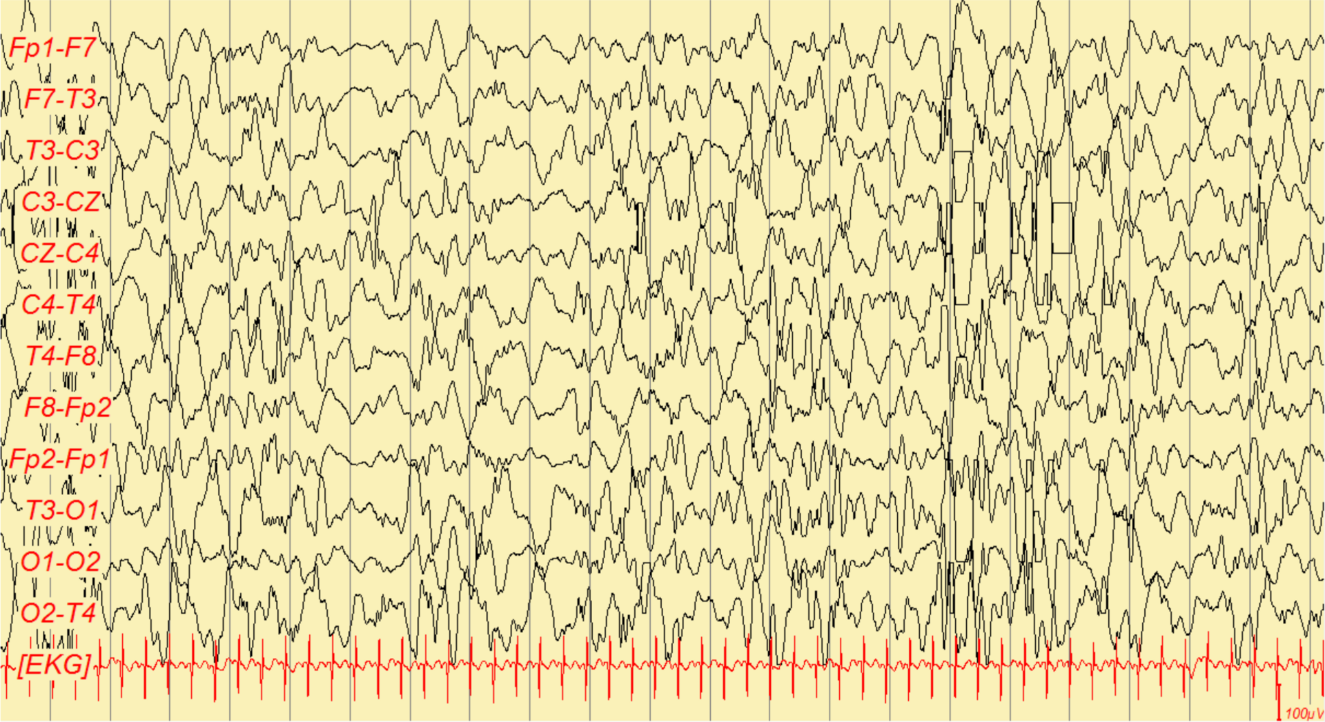
Hypsarrhythmia: chaotic, high-amplitude (>200 µV) multifocal spikes and slow waves…

Study the finding, then test the visual cue against the linked Review and Q-Book material.

View current live plate ↗
THE PLATE02 / 02
Hypsarrhythmia: chaotic, high-amplitude (>200 µV) multifocal spikes and slow waves… — source media 1
IMAGE 01 · 1920 × 1047 px · Cached rendition

Hypsarrhythmia: chaotic, high-amplitude (>200 µV) multifocal spikes and slow waves…

The QBook workbook lists this Commons file for the requested item. Commons license and dimensions were independently checked; the precise requested clinical features have not been independently confirmed.

Credit & reuse

Ralphelg · CC BY-SA 3.0 · inherited_catalog_not_refetched

License record ↗ · Media ID MED-b154ef85300b8531c8d1
Hypsarrhythmia: chaotic, high-amplitude (>200 µV) multifocal spikes and slow waves… — source media 2
IMAGE 02 · 1920 × 2188 px · Cached rendition

Hypsarrhythmia: chaotic, high-amplitude (>200 µV) multifocal spikes and slow waves…

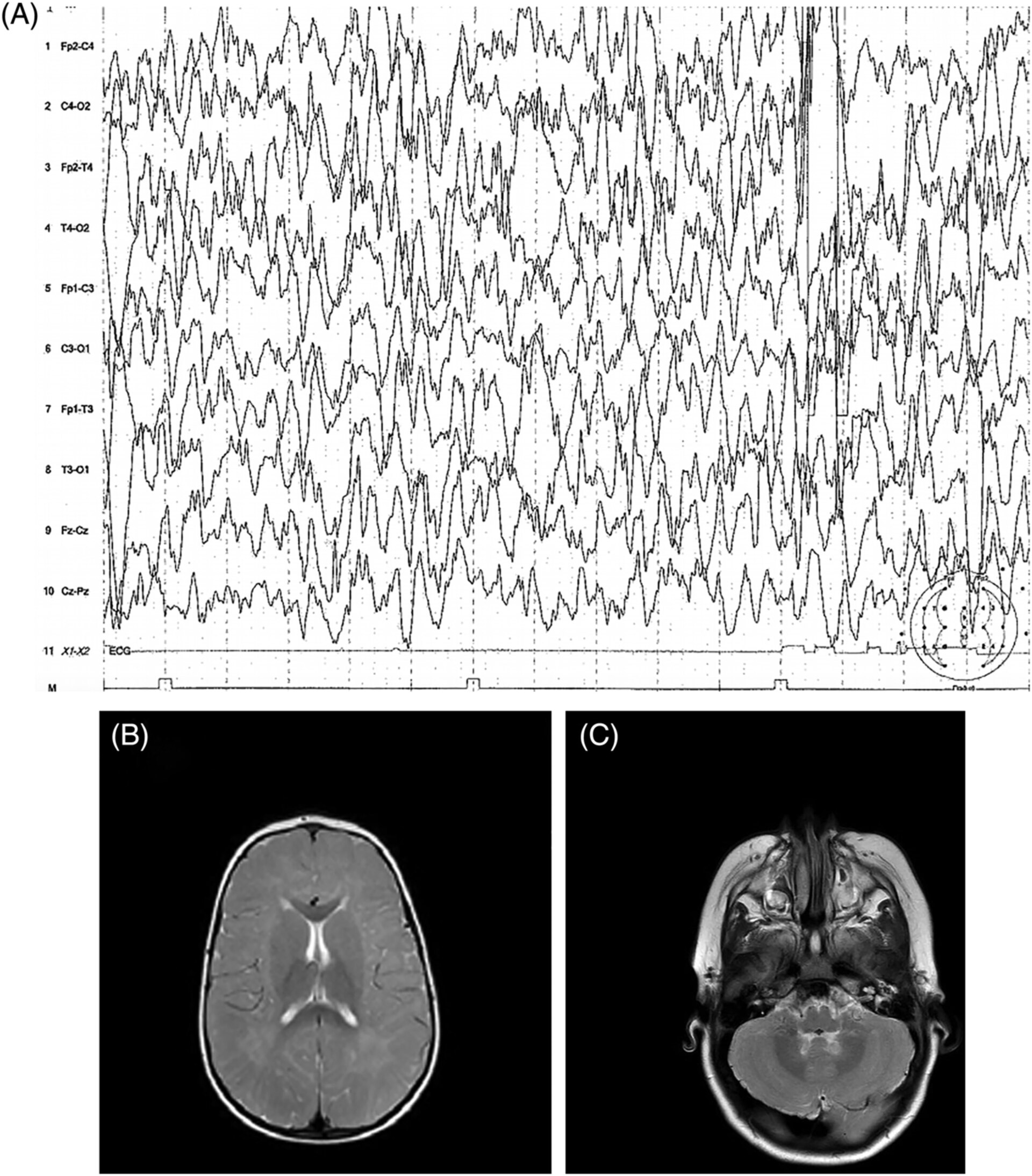
Hypsarrhythmic-like EEG activity in a child with cerebral folate deficiency. Related pattern comparator, not a classic West-syndrome case.

Credit & reuse

Maria T. Papadopoulou,Efterpi Dalpa,Michalis Portokalas,Irene Katsanika,Katerina Tirothoulaki,Martha Spilioti,Spyros Gerou,Barbara Plecko,Athanasios E. Evangeliou · CC BY 4.0 · inherited_catalog_not_refetched

License record ↗ · Media ID MED-bb2df8b89dba15093854