Skip to content
thecodexes.comIMAGE BANK
PLATE ECG-BRUGADA · 3 SOURCE FILES

Brugada pattern

Study the finding, then test the visual cue against the linked Review and Q-Book material.

View current live plate ↗
THE PLATE03 / 03
Brugada pattern — source media 1
IMAGE 01 · 4317 × 2358 px · Cached rendition

Brugada pattern

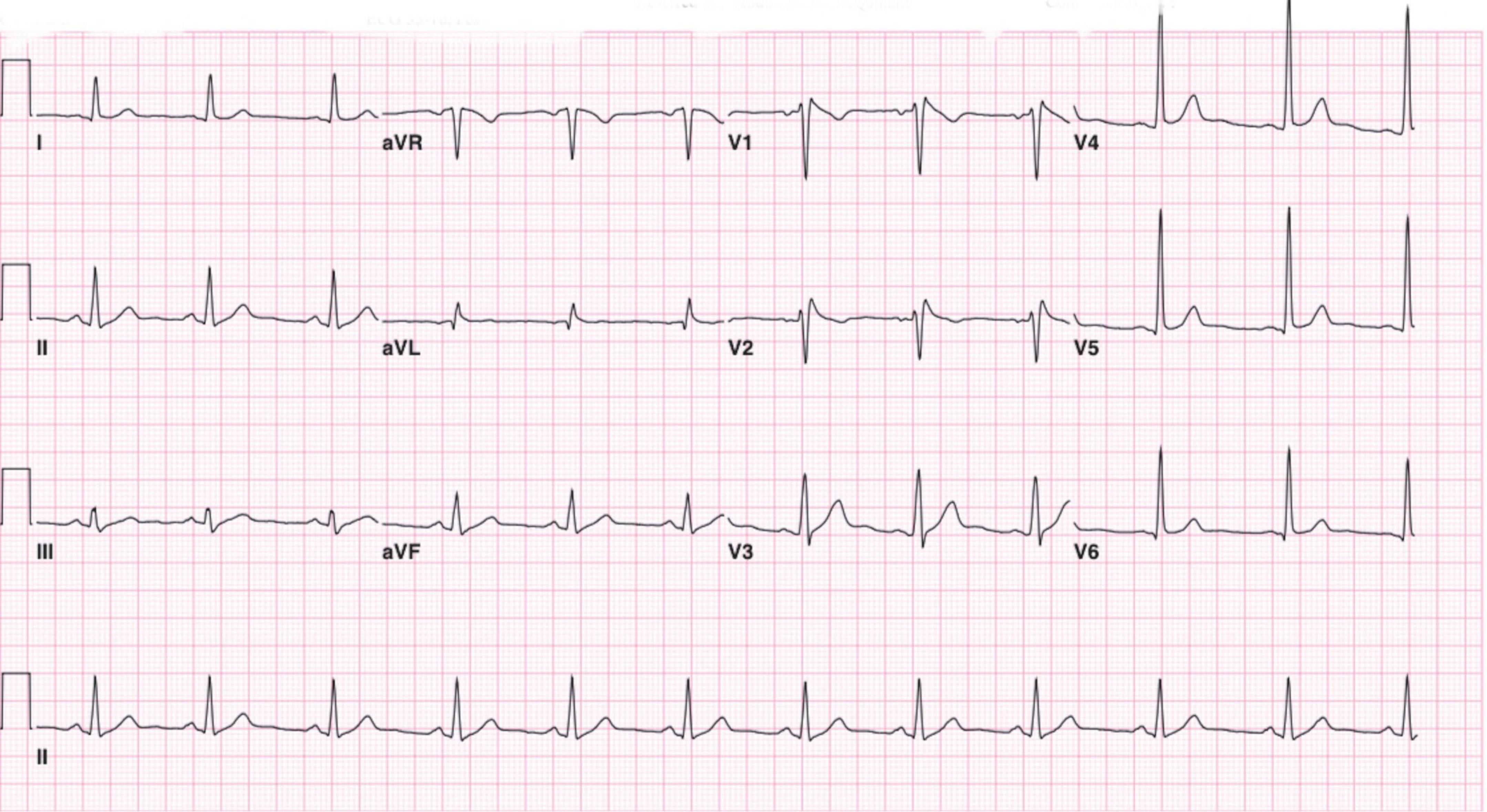
Exact high-resolution type 1 Brugada pattern, explicitly confirmed in the source caption and article text. The pattern was unmasked after lamotrigine exposure; the figure is not a normal or post-treatment tracing.

Credit & reuse

Hafiz Omer; Mohamed H. Omer; Abdulmohsen R. Alyousef; Ali M. Alzammam; Omar Ahmad; Haitham A. Alanazi · CC BY 4.0 · inherited_catalog_not_refetched

License record ↗ · Media ID MED-840f2e3cd421b1d2072e
Brugada pattern — source media 2
IMAGE 02 · 1417 × 1093 px · Cached rendition

Brugada pattern

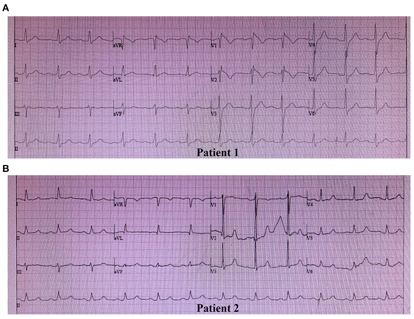
Exact high-resolution type 1 Brugada ECG in panel A, confirmed by the source caption and article text. The full publisher figure is retained because panel B is a type 3 comparison; users should not mistake panel B for the diagnostic type 1 morphology.

Credit & reuse

Meng Yuan; Yi Guo; Hong Xia; Hongbo Xu; Hao Deng; Lamei Yuan · CC BY 4.0 · inherited_catalog_not_refetched

License record ↗ · Media ID MED-6567b51dcece2d34b0dd

Brugada pattern — source media 3
IMAGE 03 · 500 × 546 px · Cached rendition

Brugada pattern

Source, credit & reuse

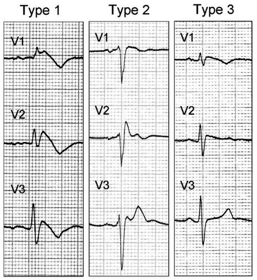
Source description identifies a type-1 Brugada ECG pattern in V1-V3; exact coved ST elevation and negative T wave remain unconfirmed.

see above · CC BY 2.0 · inherited_catalog_not_refetched

License record ↗ · Media ID MED-361e9566c30d96c902fb