IMAGE 01 · 1920 × 949 px · Cached rendition
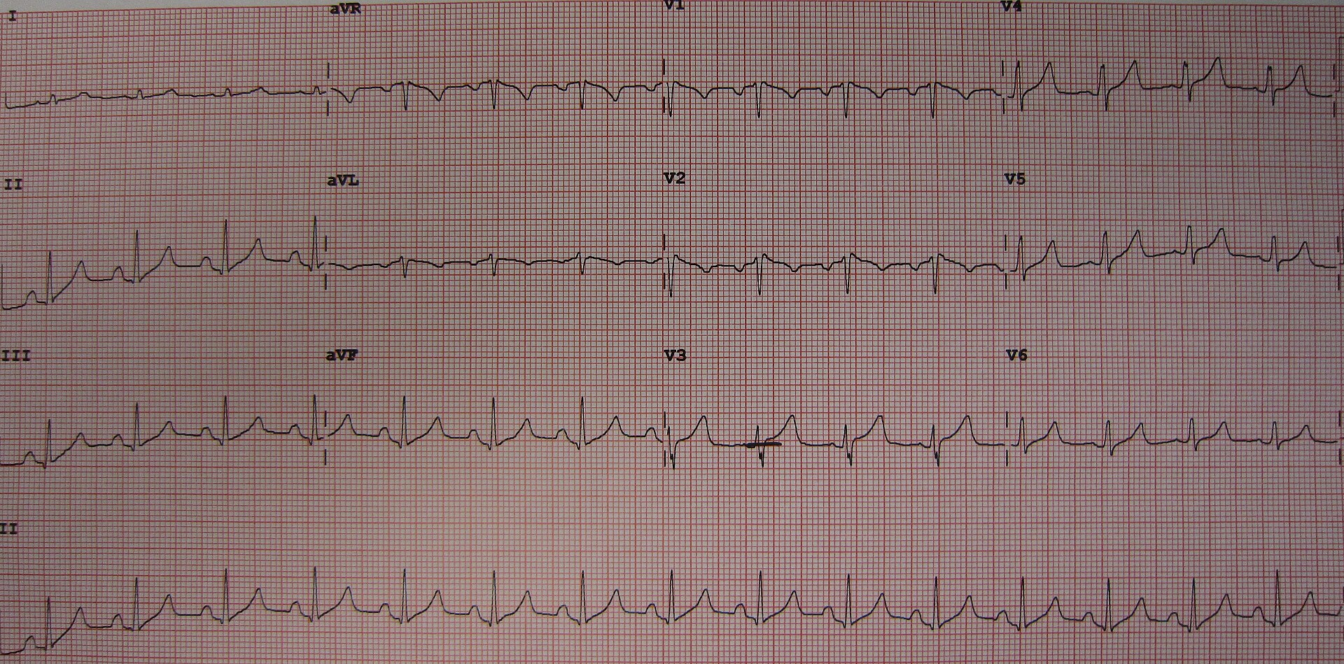
12-lead ECG with peaked narrow-based T waves in V2–V4, flattened P waves, and QRS…
The QBook workbook lists this Commons file for the requested item. Commons license and dimensions were independently checked; the precise requested clinical features have not been independently confirmed.
Credit & reuse
James Heilman, MD · CC BY-SA 3.0 · inherited_catalog_not_refetched
License record ↗ · Media ID MED-a4c211ff3dde26a1b02f