Skip to content
thecodexes.comIMAGE BANK
PLATE CXR-SARCOID · 2 SOURCE FILES

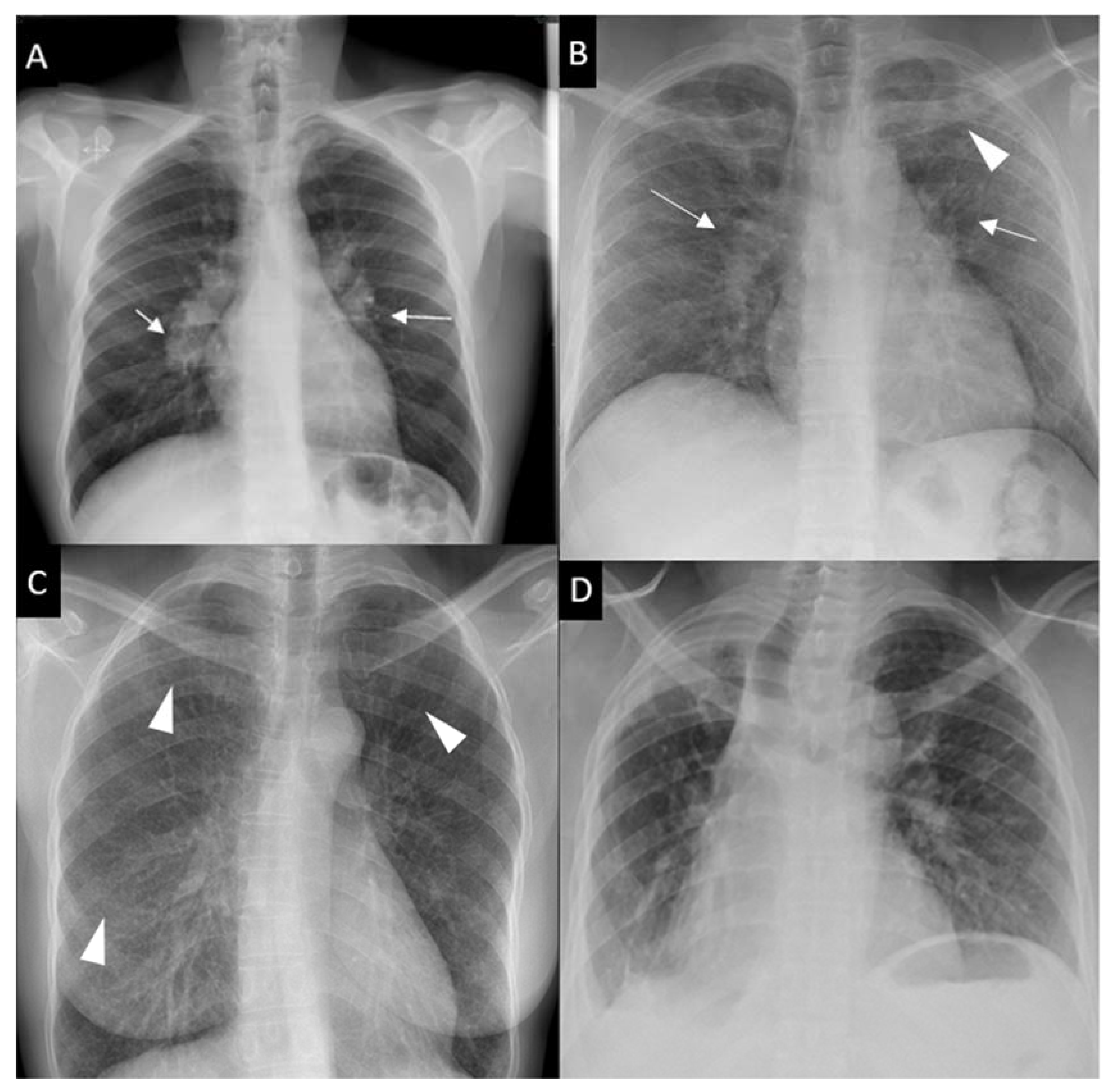
Sarcoidosis hilar adenopathy

Study the finding, then test the visual cue against the linked Review and Q-Book material.

View current live plate ↗
THE PLATE02 / 02
Sarcoidosis hilar adenopathy — source media 1
IMAGE 01 · 3470 × 3408 px · Cached rendition

Sarcoidosis hilar adenopathy

Exact disease and modality match. The composite contains the full Scadding chest-radiograph spectrum, including the hallmark bilateral hilar lymphadenopathy. It is one source figure rather than independent patient images for every stage.

Credit & reuse

Pascal Sève; Yves Pacheco; François Durupt; Yvan Jamilloux; Mathieu Gerfaud-Valentin; Sylvie Isaac; Loïc Boussel; Alain Calender; Géraldine Androdias; Dominique Valeyre; Thomas El Jammal · CC BY 4.0 · inherited_catalog_not_refetched

License record ↗ · Media ID MED-f830a059fc57d00acd5d
Sarcoidosis hilar adenopathy — source media 2
IMAGE 02 · 500 × 437 px · Cached rendition

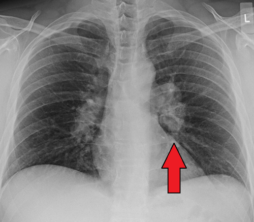
Sarcoidosis hilar adenopathy

Source, credit & reuse

Source Source description identifies sarcoid hilar adenopathy, and visual review confirms a frontal chest radiograph with bilateral hilar enlargement; exact stage remains unconfirmed.

James Heilman, MD · CC BY-SA 4.0 · inherited_catalog_not_refetched

License record ↗ · Media ID MED-17db55312f866b193860