Skip to content
thecodexes.comIMAGE BANK
PLATE CT-RENAL-STONE · 2 SOURCE FILES

Ureteral stone CT

Study the finding, then test the visual cue against the linked Review and Q-Book material.

View current live plate ↗
THE PLATE02 / 02
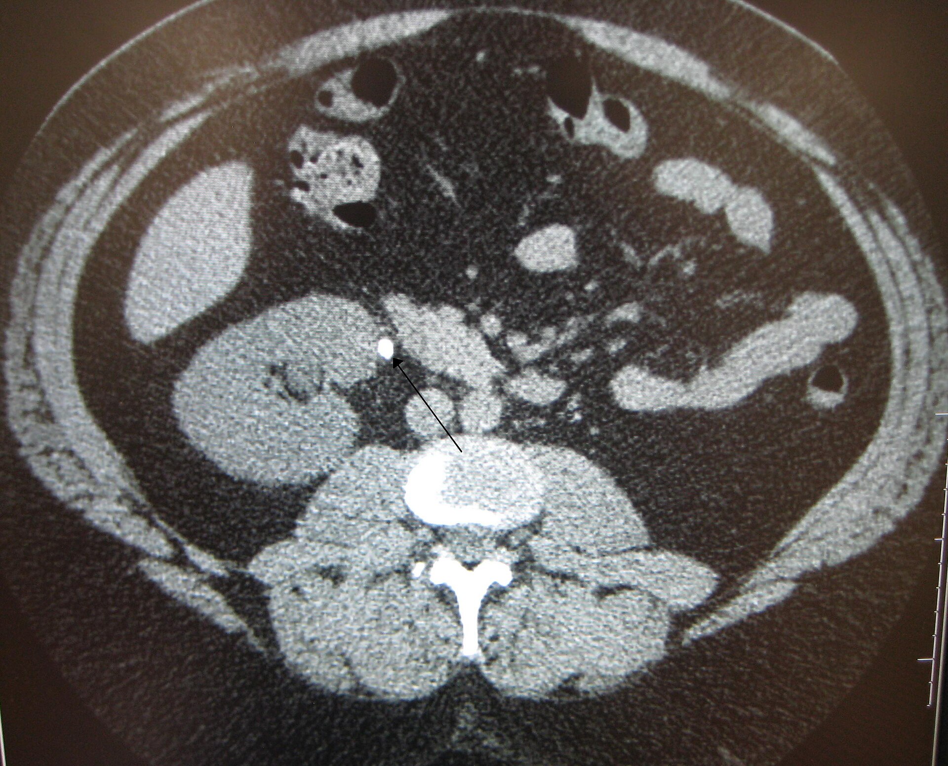
Ureteral stone CT — source media 1
IMAGE 01 · 1920 × 1552 px · Cached rendition

Ureteral stone CT

Source, credit & reuse

Source description identifies noncontrast abdominal CT with a proximal ureteric stone causing obstruction and hydronephrosis.

James Heilman, MD · CC BY-SA 3.0 · inherited_catalog_not_refetched

License record ↗ · Media ID MED-5a054b1d74a9254ac8df
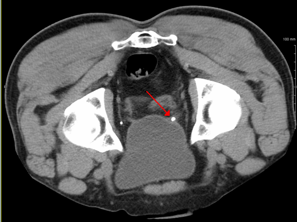
Ureteral stone CT — source media 2
IMAGE 02 · 960 × 716 px · Cached rendition

Ureteral stone CT

Stone diagnosis and CT modality/category are explicit; noncontrast technique, laterality, and hydronephrosis are unconfirmed.

Credit & reuse

James Heilman, MD · CC BY-SA 3.0 · inherited_catalog_not_refetched

License record ↗ · Media ID MED-396ceade624cea8b4529