Skip to content
thecodexes.comIMAGE BANK
PLATE CT-PE · 3 SOURCE FILES

Pulmonary embolism

Study the finding, then test the visual cue against the linked Review and Q-Book material.

View current live plate ↗
THE PLATE03 / 03
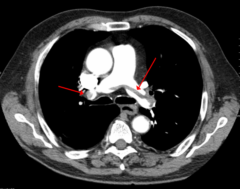
Pulmonary embolism — source media 1
IMAGE 01 · 960 × 756 px · Cached rendition

Pulmonary embolism

Diagnosis and CT topic are explicit; exact requested filling-defect morphology beyond saddle location is not separately confirmed.

Credit & reuse

James Heilman, MD · CC BY-SA 3.0 · inherited_catalog_not_refetched

License record ↗ · Media ID MED-5b851fa4e1421f14d12d
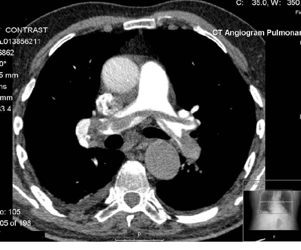
Pulmonary embolism — source media 2
IMAGE 02 · 960 × 799 px · Cached rendition

Pulmonary embolism

Diagnosis and CT modality are explicit; exact vessel extent and ancillary findings are unconfirmed.

Credit & reuse

Glitzy queen00 at English Wikipedia · Public domain · inherited_catalog_not_refetched

License record ↗ · Media ID MED-5075e0d2e167c82d86a8

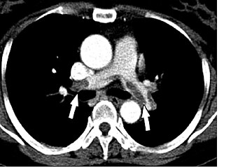
Pulmonary embolism — source media 3
IMAGE 03 · 330 × 240 px · Cached rendition

Pulmonary embolism

Source, credit & reuse

Source description identifies spiral chest CT with multiple filling defects from pulmonary emboli; exact saddle distribution remains unconfirmed.

Walter Serra, Giuseppe De Iaco, Claudio Reverberi and Tiziano Gherli · CC BY 2.0 · inherited_catalog_not_refetched

License record ↗ · Media ID MED-3a9bcd27a7ed6599ef60