Skip to content
thecodexes.comIMAGE BANK
PLATE CT-MCA-STROKE · 3 SOURCE FILES

MCA stroke CT

Study the finding, then test the visual cue against the linked Review and Q-Book material.

View current live plate ↗
THE PLATE03 / 03
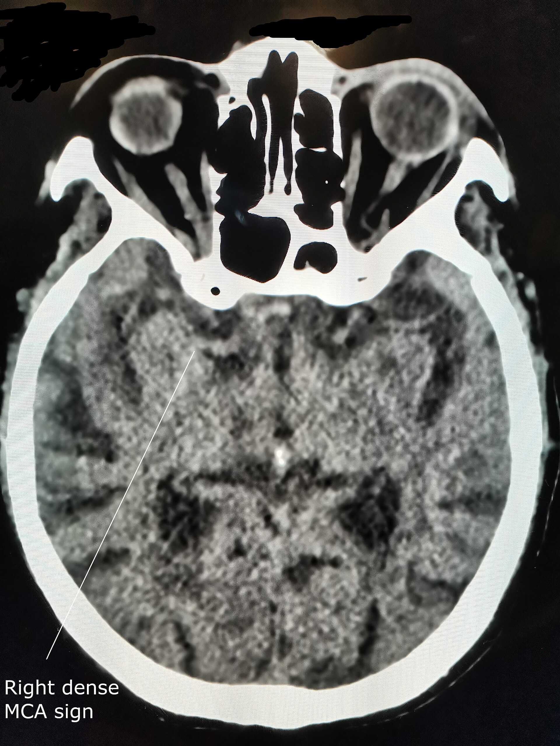
MCA stroke CT — source media 1
IMAGE 01 · 1920 × 2560 px · Cached rendition

MCA stroke CT

Source, credit & reuse

Source description explicitly identifies brain CT demonstrating a dense middle-cerebral-artery or M2 dot sign in acute MCA ischemia; early gray-white loss remains unconfirmed.

Cerevisae · CC BY-SA 4.0 · inherited_catalog_not_refetched

License record ↗ · Media ID MED-5ddb84a64b33a6ae4aad
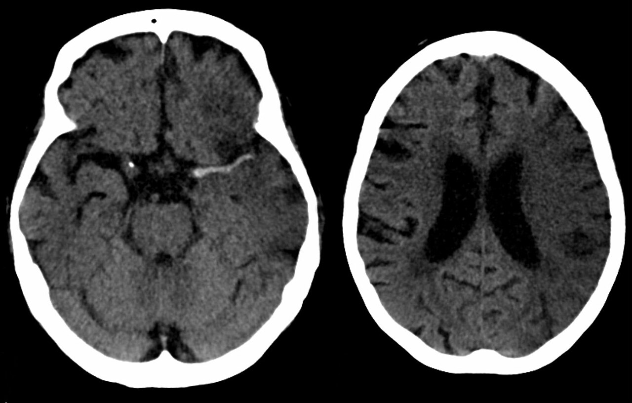
MCA stroke CT — source media 2
IMAGE 02 · 1280 × 816 px · Cached rendition

MCA stroke CT

Source, credit & reuse

Source description explicitly identifies brain CT demonstrating a dense middle-cerebral-artery or M2 dot sign in acute MCA ischemia; early gray-white loss remains unconfirmed.

Hellerhoff · CC BY-SA 3.0 · inherited_catalog_not_refetched

License record ↗ · Media ID MED-93f96d5b7c2debe22eae

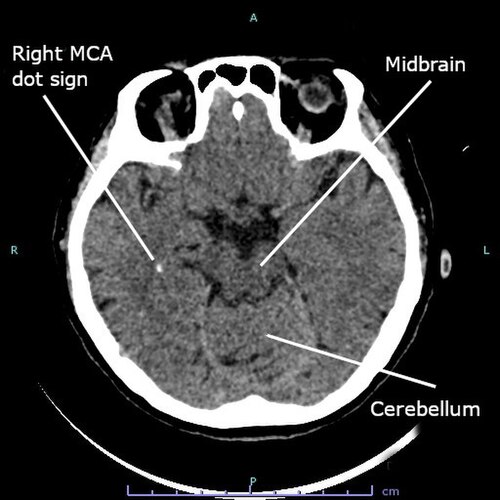
MCA stroke CT — source media 3
IMAGE 03 · 500 × 500 px · Cached rendition

MCA stroke CT

Source, credit & reuse

Source description explicitly identifies brain CT demonstrating a dense middle-cerebral-artery or M2 dot sign in acute MCA ischemia; early gray-white loss remains unconfirmed.

Cerevisae · CC BY-SA 4.0 · inherited_catalog_not_refetched

License record ↗ · Media ID MED-0f8560db08313b6cd5ae