Skip to content
thecodexes.comIMAGE BANK
PLATE CT-APPENDICITIS · 3 SOURCE FILES

Acute appendicitis

Study the finding, then test the visual cue against the linked Review and Q-Book material.

View current live plate ↗
THE PLATE03 / 03
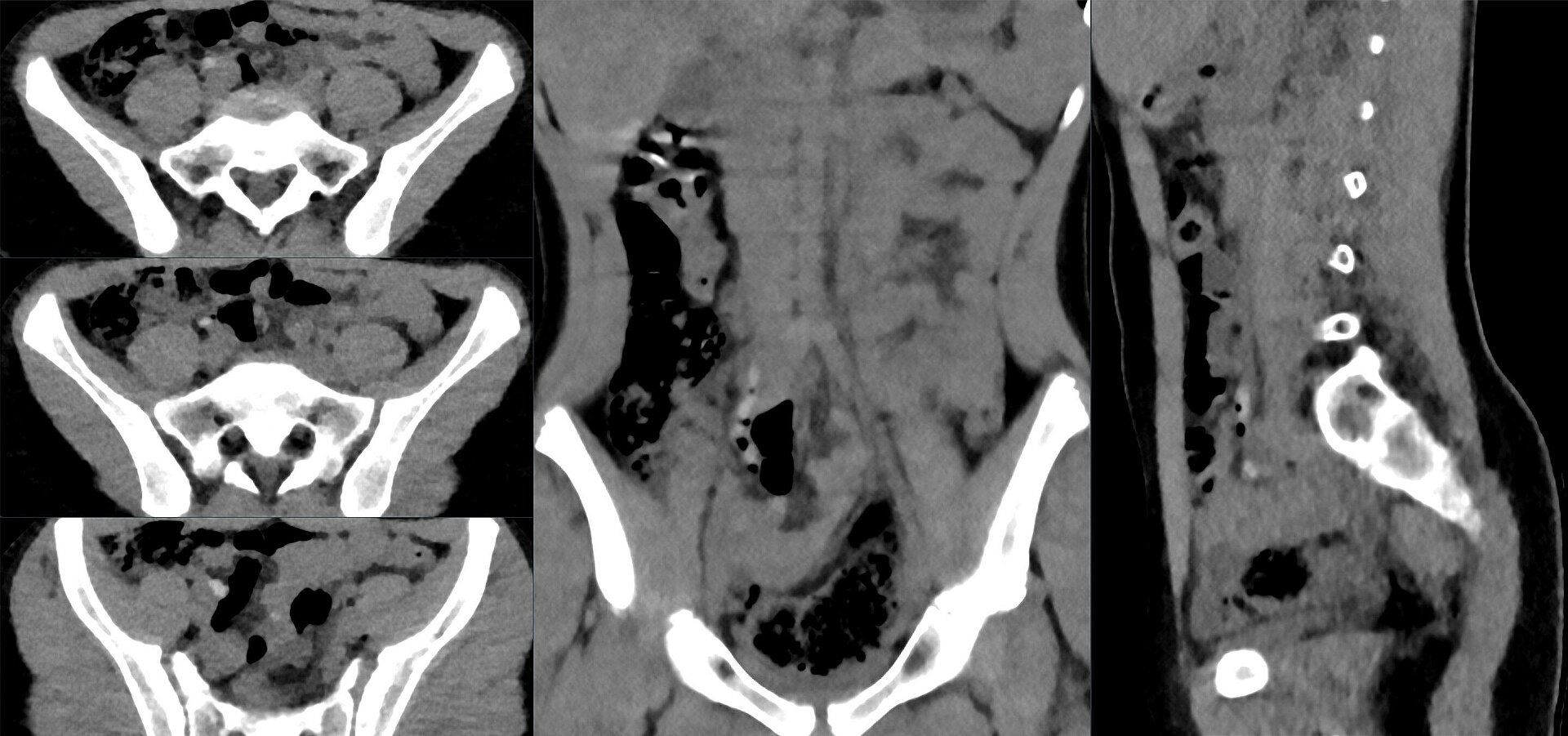
Acute appendicitis — source media 1
IMAGE 01 · 1920 × 901 px · Cached rendition

Acute appendicitis

Appendicitis topic and CT modality are explicit; diameter, wall enhancement, and periappendiceal fat stranding are unconfirmed.

Credit & reuse

Hellerhoff · CC BY-SA 4.0 · inherited_catalog_not_refetched

License record ↗ · Media ID MED-0903d70de273ee1535de
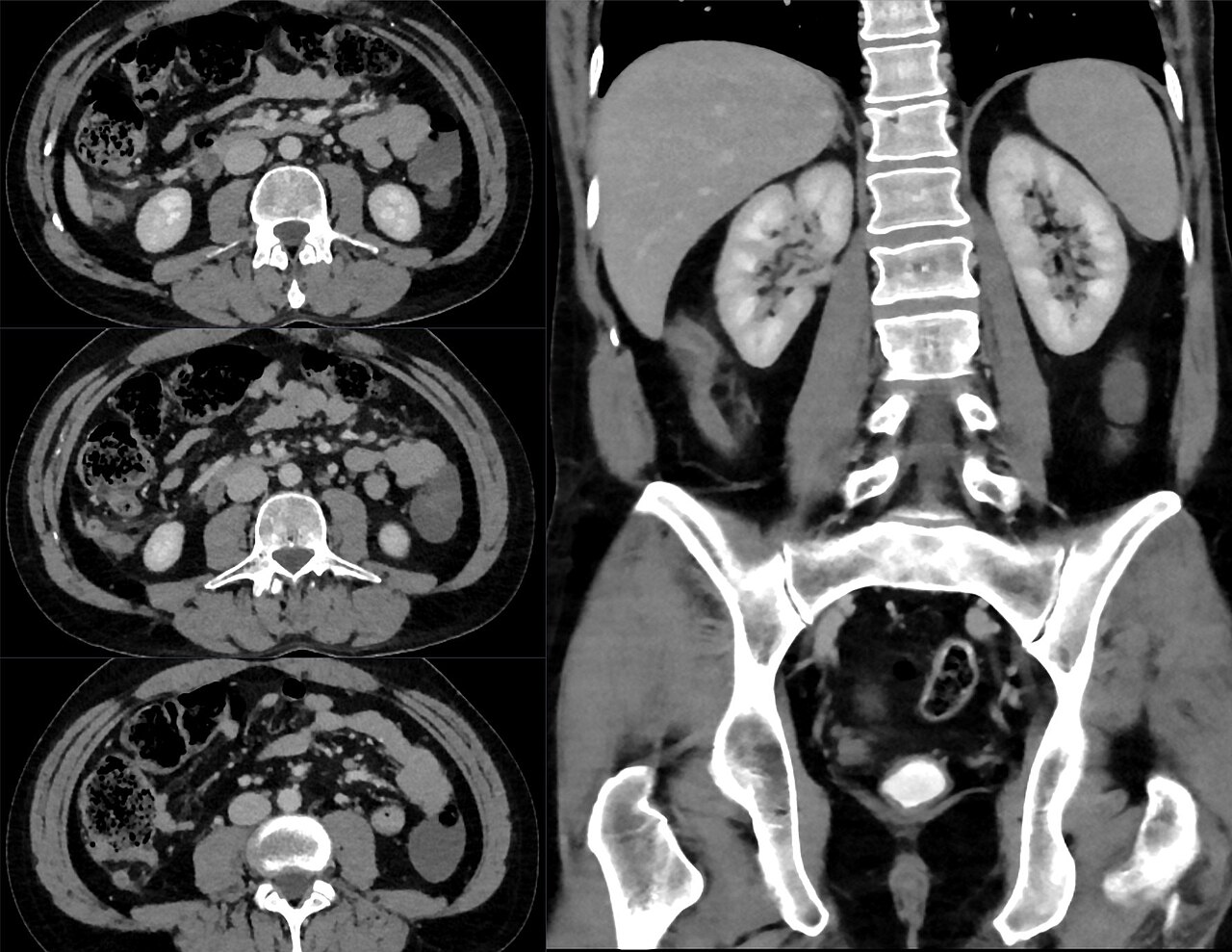
Acute appendicitis — source media 2
IMAGE 02 · 1280 × 989 px · Cached rendition

Acute appendicitis

Diagnosis and CT modality are explicit; atypical subhepatic location should be considered when teaching classic right-lower-quadrant disease.

Credit & reuse

Hellerhoff · CC BY-SA 4.0 · inherited_catalog_not_refetched

License record ↗ · Media ID MED-fa23f53c53185a69ff7a

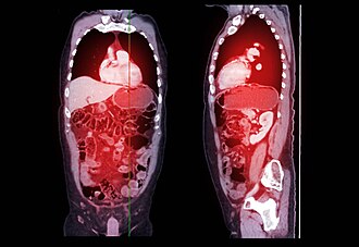
Acute appendicitis — source media 3
IMAGE 03 · 330 × 227 px · Cached rendition

Acute appendicitis

Source, credit & reuse

Source description identifies contrast-enhanced CT of acute appendicitis; dilation, enhancement, and fat stranding remain unconfirmed.

Goleisureintl · CC BY 4.0 · inherited_catalog_not_refetched

License record ↗ · Media ID MED-081419ac97e4c978ca50