Skip to content
thecodexes.comIMAGE BANK
PLATE US-OVARIAN-TORSION · 2 SOURCE FILES

Ovarian torsion on ultrasound

Study the finding, then test the visual cue against the linked Review and Q-Book material.

THE PLATE02 / 02
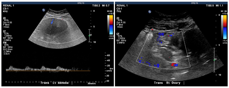
Absent left adnexal flow versus right ovary — source media 1
IMAGE 01 · 750 × 289 px · Cached rendition

Absent left adnexal flow versus right ovary

Side-by-side Doppler views show little/no detectable left adnexal vascular signal versus flow in the right ovary.

Credit & reuse

Yuchen Duan, Bryce Hoer, and Andrew Little · CC BY 4.0 · source_rechecked_this_run

License record ↗ · Media ID MED-3cf0a8461165c8a7203d
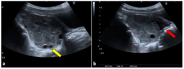
Enlarged ovary on gray-scale ultrasound — source media 2
IMAGE 02 · 729 × 275 px · Cached rendition

Enlarged ovary on gray-scale ultrasound

Two gray-scale transabdominal sonograms show enlarged, abnormally positioned ovary with peripheral follicles.

Credit & reuse

Paolo Niccolò Franco, Alejandra García-Baizán, María Aymerich, Cesare Maino, Sofia Frade-Santos, Davide Ippolito, and Milagros Otero-García · CC BY 4.0 · source_rechecked_this_run

License record ↗ · Media ID MED-141d666d85f052cea235