IMAGE 01 · 2250 × 1595 px · Browser preview; TIFF original available
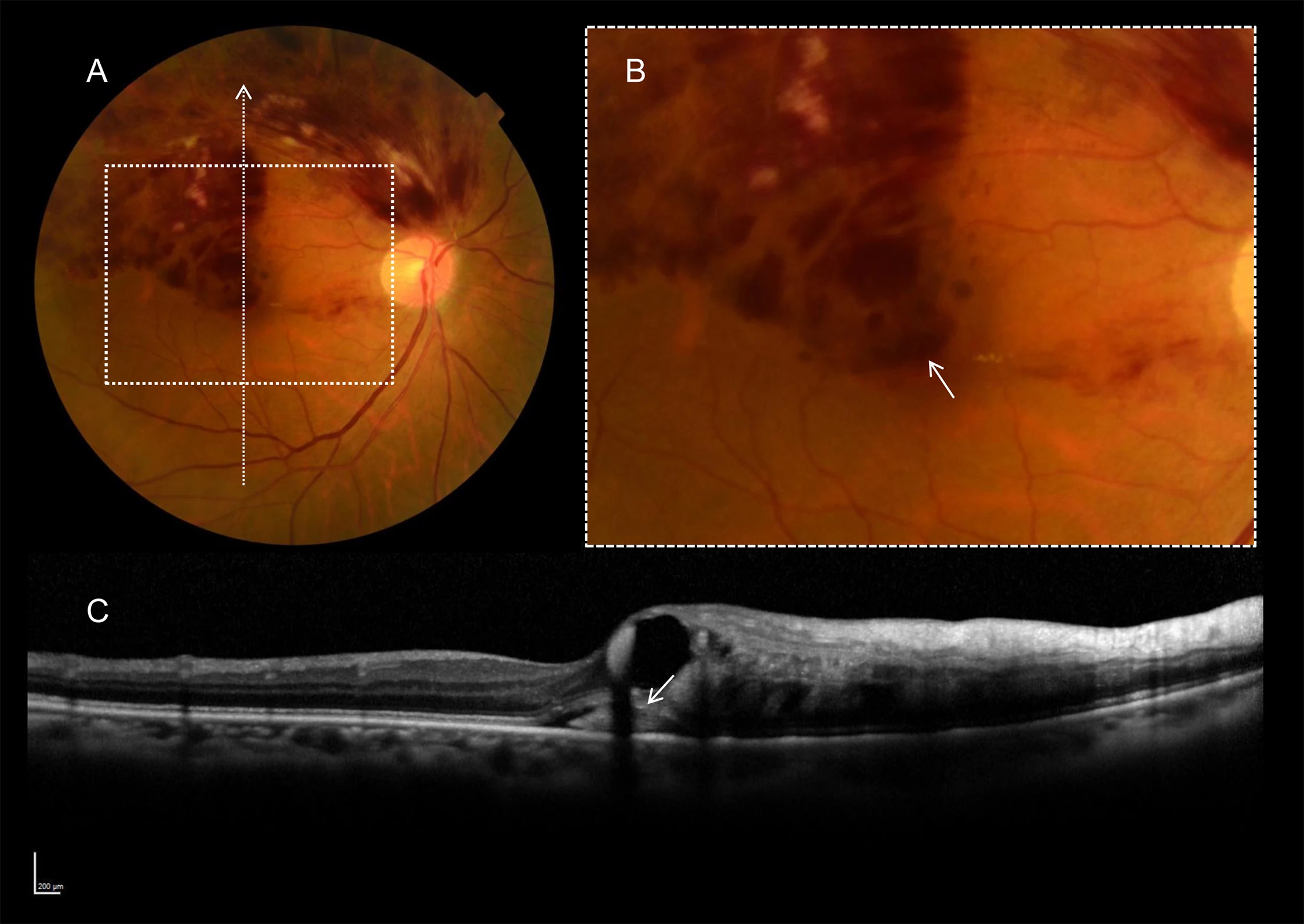
Subfoveal hemorrhage in branch retinal vein occlusion
Fundus panel shows sectoral retinal hemorrhage with macular involvement; magnified inset marks subfoveal hemorrhage, and an OCT cross section appears below.
Credit & reuse
Yuki Muraoka, Akitaka Tsujikawa, Ayako Takahashi, Yuto Iida, Tomoaki Murakami, Sotaro Ooto, Kiyoshi Suzuma, Akihito Uji, and Nagahisa Yoshimura · CC BY 4.0 · source_rechecked_this_run
License record ↗ · Media ID MED-4bdbc65904a99652c3b4